
Download
RESEARCH ARTICLE
Alpinetin suppresses cell proliferation and metastasis in osteosarcoma by inhibiting PI3K/AKT and ERK pathways
Zhenyu Cao*, Jianwu Ma, Xiaozhong Shen
Department of Orthopedics, Qinghai Provincial People’s Hospital, Xining, Qinghai Province, China
Abstract
Alpinetin, a natural flavonoid found in medicinal herbs, possesses distinct pharmacological activities, including neuroprotective, antiviral, antibacterial, lung and cardiovascular protective, hepatoprotective, antiinflammatory, and antitumor properties. Here, the role of alpinetin was investigated in osteosarcoma. Osteosarcoma cell lines (143B and U2OS) were treated with 10-, 25-, 50-, 75-, or 100-μM concentration of alpinetin. Cell proliferation was detected by cell counting kit 8 (CCK8) and colony formation assays. Western blotting and flow cytometry were used to investigate cell apoptosis. Cell metastasis was assessed by transwell assay. Administration of alpinetin reduced the viabilities of 143B and U2OS cell lines in a dosage-dependent manner, and decreased the number of colonies of 143B and U2OS cell lines. The apoptosis of 143B and U2OS cell lines was promoted by alpinetin through down-regulation of Bcl-2 and up-regulation of cleaved caspase-3 and Bax. Alpinetin inhibited invasion and migration of 143B and U2OS cell lines through up-regulation of epithelial biomarkers, E-cadherin, and zonula occludens-1 (ZO-1), and down-regulation of mesenchymal biomarkers, Vimentin, and N-cadherin. Alpinetin also reduced the phosphorylation of extracellular signal-regulated kinase (ERK), Ak strain transforming (AKT), and phosphoinositide 3-kinases (PI3K) in 143B and U2OS cell lines. Alpinetin inhibited cell proliferation and metastasis in osteosarcoma through inactivation of PI3K/AKT and ERK pathways, providing potential treatment option for treatment of cancer.
Key words: alpinetin, proliferation, metastasis, epithelial to mesenchymal transition, osteosarcoma, PI3K/AKT, ERK
*Corresponding Author: Zhenyu Cao, Department of Orthopedics, Qinghai Provincial People’s Hospital, No. 2 Gonghe Road, Chengdong District, Xining City, Qinghai Province 810000, China. Email: [email protected]
Received: 3 March 2022; Accepted: 6 April 2022; Published: 5 May 2022
© 2022 Codon Publications
This is an Open Access article distributed under the terms of the Creative Commons Attribution-NonCommercial-ShareAlike 4.0 International (CC BY-NC-SA 4.0). License (http://creativecommons.org/licenses/by-nc-sa/4.0/)
Introduction
Osteosarcoma is the most common primary bone tumor observed in adolescents and children (Dengra et al., 2012). Surgery and adjuvant chemotherapy have improved the 5-year overall survival rate of patients with osteosarcoma from 20% to 75% (Abdelgawad et al., 2022). However, development of metastasis, recurrence, and chemoresistance reduces the prognosis of osteosarcoma (He et al., 2014). Therefore, new therapeutic agents to suppress the metastasis of osteosarcoma are urgently required to optimize treatment strategies.
Natural flavonoid-rich herbs, including Urtica dioica (stinging nettle), ameliorated cognitive dysfunction in streptozotocin-induced diabetic mice via reduction of neuroinflammation and hippocampal oxidative stress (Keshvari et al., 2020; Rahmati et al., 2021). Alpinetin, a natural flavonoid found in medicinal herbs, possesses distinct pharmacological activities, including neuroprotective, antiviral, antibacterial, lung and cardiovascular protective, hepatoprotective, antiinflammatory, and antitumor properties (Zhao et al., 2022). In tumors, alpinetin promoted cell apoptosis, inhibited cell invasion and metastasis, and induced cell cycle arrest in breast, lung, and tongue squamous carcinoma, glioma, hepatic, pancreatic, gastric, colon, and cervical cancers (Zhao et al., 2022). Moreover, alpinetin also ameliorated cancer cachexia of Lewis lung carcinoma through activation of peroxisome proliferator-activated receptor-γ (Zhang et al., 2021). The proliferation and invasion of ovarian cancer cells were also repressed by alpinetin (Zhao et al., 2018). However, the anticancer role of alpinetin in osteosarcoma has not been reported for the time being.
Phosphoinositide 3-kinases (PI3K)/Ak strain transforming (AKT) signaling, important for cell survival (Qiu et al., 2021), was abnormally activated in osteosarcoma (Zhang et al., 2015). Activation of PI3K/AKT signaling contributed to cell proliferation, invasion, angiogenesis, metastasis, and chemoresistance of osteosarcoma (Zhang et al., 2015). Inhibition of PI3K/AKT signaling via PI3K inhibitors or natural compounds from plants was used as therapeutic strategies for osteosarcoma (Zhang et al., 2015). Alpinetin has been demonstrated to enhance chemosensitivity of lung cancer cells and suppress progression of tumor through inactivation of PI3K/AKT signaling (Wu et al., 2015). It is thus hypothesized that alpinetin might also suppress osteosarcoma cell proliferation and metastasis through inhibition of PI3K/AKT signaling. Therefore, the effects of alpinetin on cell proliferation, apoptosis, invasion, and migration of osteosarcoma were investigated in this study.
Materials and methods
Cell culture and treatment
Human B lymphoblast (IM-9) and osteosarcoma cell lines (143B and U2OS) were purchased from ATCC (Manassas, VA, USA), and grown in RPMI 1640 medium (Thermo Fisher Scientific, Waltham, MA, USA) with 10% fetal bovine serum (Sigma-Aldrich, St. Louis, MO, USA). Cells were treated with 10-, 25-, 50-, 75-, and 100-μM alpinetin (Sigma-Aldrich) for 24 h, and then subjected to functional assays.
Cell proliferation assays
IM-9 and osteosarcoma cell lines were seeded into 96-well plates, and incubated with different concentrations of alpinetin for 24 h. Cells were then treated with cell counting kit 8 (CCK8) solution (Beyotime, Beijing, China) for another 2 h. Absorbance at 490 nm was measured by a microplate reader (Thermo Fisher Scientific).
In order to detect cell proliferation, osteosarcoma cells were seeded into 6-well plates, and incubated with different concentrations of alpinetin for 24 h. Cells were then grown in RPMI 1640 medium for 10 days. Cell colonies were fixed in methanol, stained with 0.1% crystal violet (Sigma-Aldrich), and photographed under light microscope (Olympus, Tokyo, Japan).
Cell apoptosis assay
Osteosarcoma cells were performed with trypsin digestion, harvested and resuspended in binding buffer of BD Cycletest™ Plus DNA Reagent Kit (BD Biosciences, San Jose, CA). Cells were stained with propidium oxide and fluorescein isothiocyanate-conjugated annexin V, and analyzed under FACS flow cytometer (Life Technologies, Darmstadt, Germany).
Transwell assays
Osteosarcoma cells in serum-free RPMI 1640 medium were plated into upper Transwell insert chamber (Corning Incorporated, Corning, NY, USA). RPMI 1640 medium with 15% fetal bovine serum was plated into the lower chamber. Invasive cells in the lower chamber were stained with crystal violet and observed under microscope (Olympus) after 24 h.
In order to assess cell invasion, osteosarcoma cells in serum-free medium were also plated into Matrigel-coated upper chambers and subjected to the same protocol.
Western blotting
Osteosarcoma cells were lysed in radioimmunoprecipitation assay (RIPA) buffer (Beyotime), and the isolated proteins were then separated by 10% sodium dodecyl sulfate–polyacrylamide gel electrophoresis (SDS-PAGE). Proteins were transferred onto nitrocellulose membranes, and the membranes were blocked in 5% bovine serum albumin. Membranes were probed with specific antibodies: anti-PCNA and anti-β-actin (1:2,000), anti-cleaved caspase-3 and anti-caspase-3 (1:2,500), anti-Bax and anti-Bcl-2 (1:3,000), anti-Vimentin and anti-N-cadherin (1:3,500), anti-E-cadherin and anti-ZO-1 (1:4,000), anti-p-extracellular signal-regulated kinase (ERK) and anti-ERK (1:4,500), anti-p-AKT and anti-AKT (1:5,000), and anti-p-PI3K and anti-PI3K (1:5,500). The membranes were then washed and incubated with horseradish peroxidase-conjugated secondary antibody (1:5,000). Immunoreactivities were visualized using enhanced chemiluminescence (Sigma-Aldrich), and the blots of proteins were quantified by Image J using β-actin as an internal reference. All the antibodies were obtained from Abcam (Cambridge, MA, USA).
Statistical analysis
All data with at least triple replicates were expressed as mean ± standard error of mean (SEM), and analyzed by Student’s t-test or one-way analysis of variance (ANOVA) with post-hoc analysis using the SPSS software. The normality and homogeneity of data were investigated by Shapiro–Wilk and Levene’s tests, respectively. P < 0.05 was considered as statistically significant.
Results
Alpinetin reduced cell proliferation of osteosarcoma
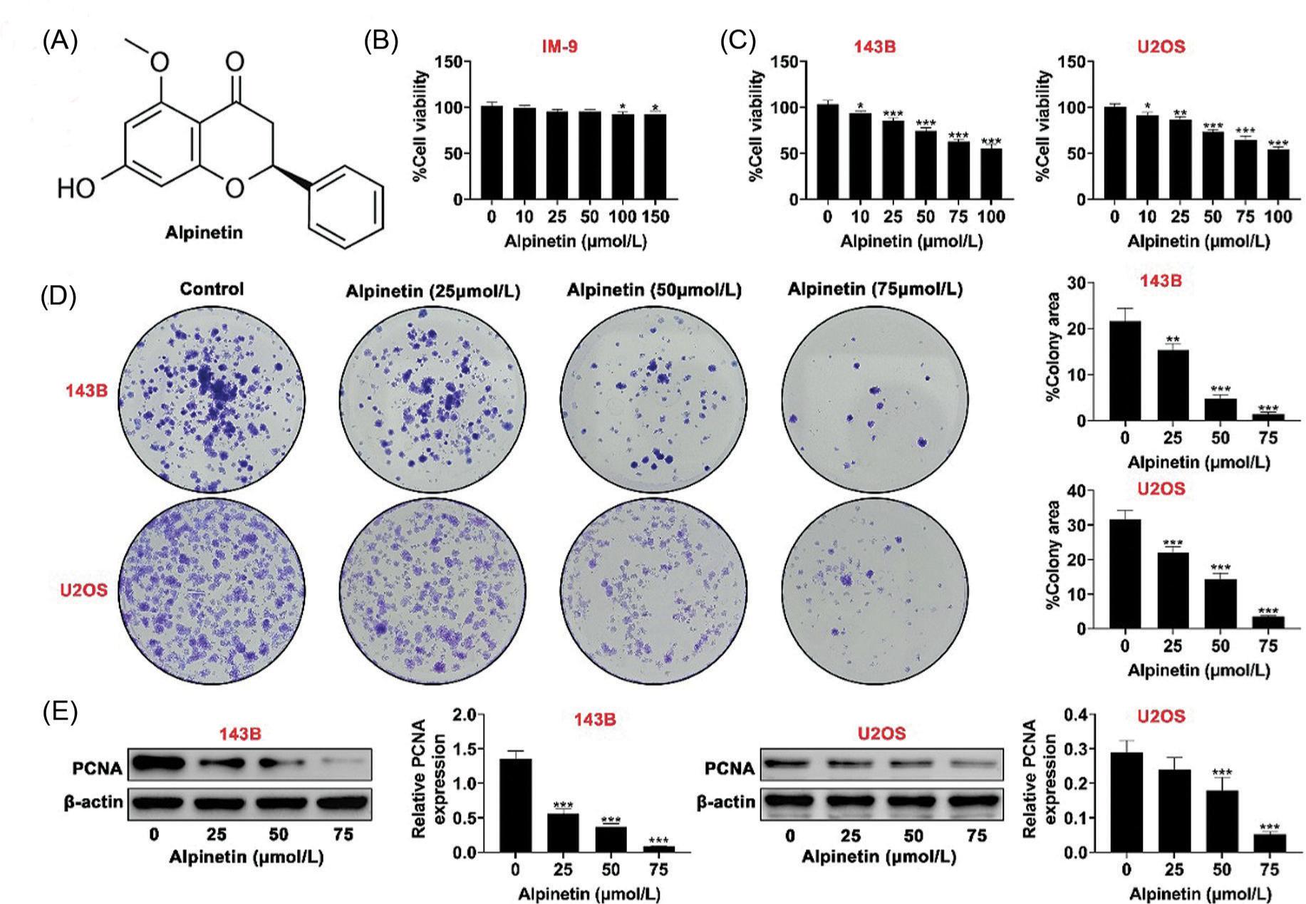
In order to investigate the cytotoxicity of alpinetin in B lymphoblast, IM-9 cell line was treated with 10-, 25-, 50-, 75-, or 100-μM alpinetin (Figure 1A). Treatment with alpinetin below 50 μM did not affect the viability of IM-9 cell (Figure 1B), and 75- or 100-μM alpinetin reduced no more than 10% of viability of IM-9 cell (Figure 1B). However, treatment with alpinetin decreased the viabilities of osteosarcoma cell lines (143B and U2OS) in a dosage-dependent manner (Figure 1C). Number of colonies in 143B and U2OS cell lines were reduced by alpinetin (Figure 1D) through down-regulation of proliferation-related biomarker, proliferating cell nuclear antigen (PCNA) (Figure 1E), suggesting the antiproliferative effect of alpinetin on osteosarcoma.
Figure 1. Alpinetin reduced cell proliferation of osteosarcoma. (A) Chemical structure of alpinetin. (B) Treatment with alpinetin reduced the viability of IM-9 cell line, detected by CCK8 assay. (C) Treatment with alpinetin decreased the viabilities of 143B and U2OS cell lines in a dosage-dependent manner, detected by CCK8 assay. (D) Treatment with alpinetin decreased the proliferation of 143B and U2OS cell lines, detected by colony formation assay. (E) Treatment with alpinetin decreased the protein expression of PCNA in 143B and U2OS cell lines. *P < 0.05, **P < 0.01, ***P < 0.001.
Alpinetin promoted cell apoptosis of osteosarcoma
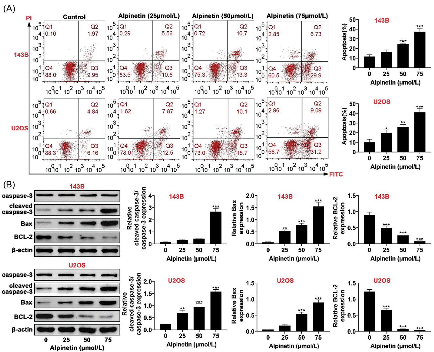
In order to investigate the role of alpinetin in cell apoptosis of osteosarcoma, flow cytometry was performed, and the results demonstrated that alpinetin promoted the apoptosis of 143B and U2OS cell lines in a dosage- dependent manner (Figure 2A). The protein expression of pro-survival biomarker, Bcl-2, was down-regulated in 143B and U2OS cell lines by alpinetin (Figure 2B). However, the expression of apoptotic biomarkers, cleaved caspase-3 and Bax, were up-regulated in 143B and U2OS cell lines by alpinetin (Figure 2B), demonstrating the pro-apoptotic effect of alpinetin on osteosarcoma.
Figure 2. Alpinetin promoted cell apoptosis of osteosarcoma. (A) Treatment with alpinetin promoted the apoptosis of 143B and U2OS cell lines in a dosage-dependent manner, detected by flow cytometry assay. (B) Treatment with alpinetin down-regulated Bcl-2, up-regulated cleaved caspase-3 and Bax of 143B and U2OS cell lines in a dosage-dependent manner. **P < 0.01, ***P < 0.001.
Alpinetin reduced cell metastasis of osteosarcoma
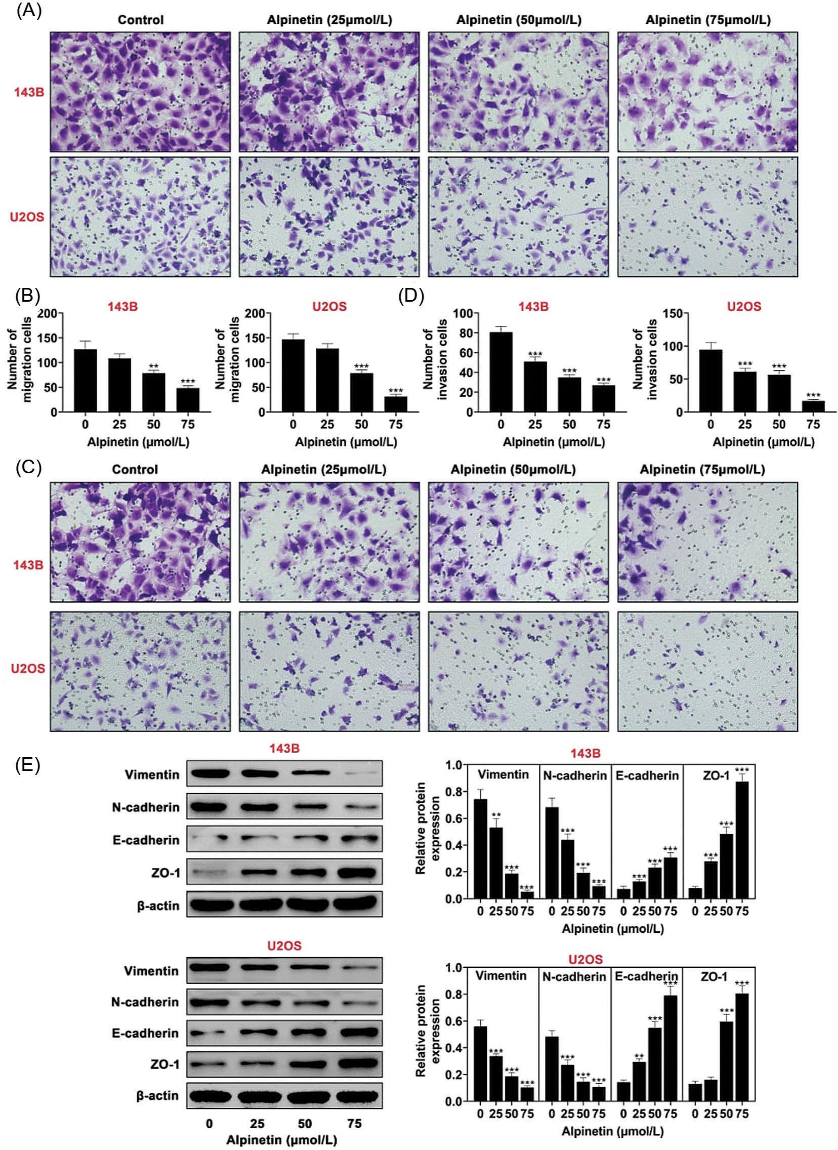
In order to investigate the role of alpinetin in cell metastasis of osteosarcoma, transwell assays were performed; the results indicated that alpinetin inhibited the migration of 143B and U2OS cell lines (Figure 3A) and decreased the number of invasive cells of 143B and U2OS cell lines in a dosage-dependent manner (Figure 3B), suggesting that the migration and invasion of osteosarcoma cells were suppressed by alpinetin. The protein expressions of mesenchymal biomarkers, vimentin and N-cadherin, were decreased, while that of epithelial biomarkers, E-cadherin and ZO-1, were increased in 143B and U2OS cell lines by alpinetin in a dosage-dependent manner (Figure 3C), indicating that alpinetin suppressed epithelial to mesenchymal transition in osteosarcoma.
Figure 3. Alpinetin reduced cell metastasis of osteosarcoma. (A) Alpinetin inhibited cell migration of 143B and U2OS cell lines in a dosage-dependent manner detected by transwell assay. (B) Alpinetin inhibited cell invasion of 143B and U2OS in a dosage-dependent manner detected by transwell assay. (C) Alpinetin inhibited decreased protein expression of vimentin and N-cadherin, increased E-cadherin and ZO-1 of 143B and U2OS cell lines in a dosage-dependent manner. **P < 0.01, ***P < 0.001.
Alpinetin suppressed PI3K/AKT and ERK signaling in osteosarcoma
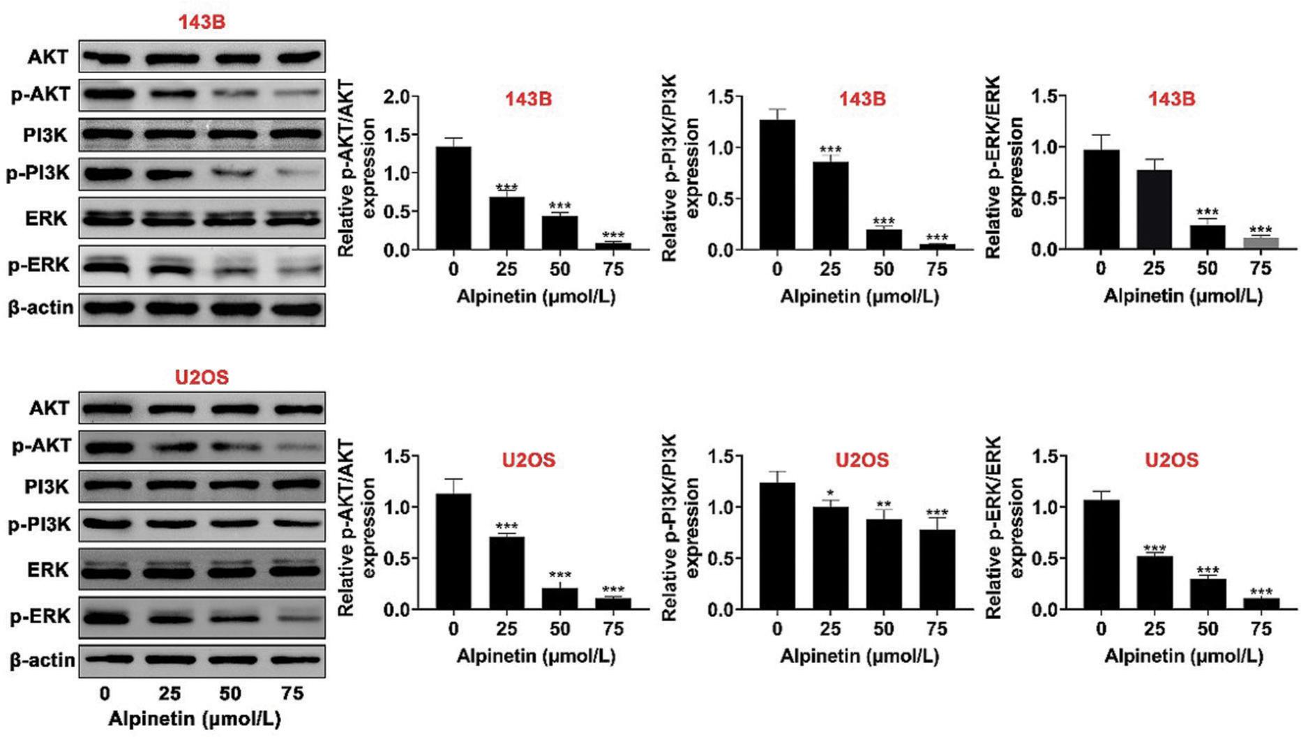
In order to clarify the mechanism underlying the effects of alpinetin on mediating progression of osteosarcoma, Western blotting was performed. The results demonstrated that the protein expressions of ERK in 143B and U2OS cell lines were not affected by alpinetin (Figure 4), while p-ERK was reduced by alpinetin in a dosage-dependent manner (Figure 4). Moreover, alpinetin down-regulated p-AKT and p-PI3K in 143B and U2OS cell lines (Figure 4), revealing that alpinetin repressed the activation of PI3K/AKT and ERK signaling in osteosarcoma.
Figure 4. Alpinetin suppressed PI3K/AKT and ERK signaling in osteosarcoma. Alpinetin down-regulated p-ERK, p-AKT, and p-PI3K of 143B and U2OS cell lines in a dosage-dependent manner. *P < 0.05, **P < 0.01, ***P < 0.001.
Discussion
Naturally, flavonoids in herbs depict beneficial effects on various diseases, and have anticarcinogenic properties through modulation of cellular enzymes (Panche et al., 2016). For example, taxifolin, a plant flavonoid, suppresses cell proliferation and metastasis of osteosarcoma (Chen et al., 2018). The antitumor activity of alpinetin has been widely investigated in various carcinoma cells (Zhao et al., 2022). The present study found that alpinetin enhanced cell apoptosis, reduced cell proliferation, invasion, and migration of osteosarcoma.
First, functional assays in this study demonstrated that alpinetin decreased cell viability and reduced cell proliferation in osteosarcoma. PCNA, as a regulator of cell cycle, was up-regulated in patients with osteosarcoma, and predicted poor prognosis (Wang et al., 2017). Alpinetin exerted antiproliferative effect against osteosarcoma through down-regulation of PCNA. Pro-apoptotic strategies have been widely used in the treatment of osteosarcoma (Li et al., 2016). Cell apoptosis of osteosarcoma was enhanced by alpinetin through down-regulation of Bcl-2, and up-regulation of cleaved caspase-3 and Bax. Therefore, alpinetin could inhibit the progression of osteosarcoma through antiproliferative and pro-apoptotic properties.
Metastatic pattern contributed to poor prognosis in patients with osteosarcoma, and inhibition of cell metastasis in osteosarcoma was regarded as a promising strategy for the treatment of osteosarcoma (Sheng et al., 2021). Transition of epithelial to mesenchymal was implicated in the pathogenesis of metastasis in osteosarcoma (Yang et al., 2013). Promotion of epithelial to mesenchymal transition also attributed to osteosarcoma cell metastasis (Zhu et al., 2020). Here, alpinetin suppressed cell migration and invasion in osteosarcoma. Moreover, alpinetin reduced the protein expressions of mesenchymal biomarkers, including vimentin and N-cadherin, while enhanced protein expressions of epithelial biomarkers, including E-cadherin and ZO-1, to suppress transition of epithelial to mesenchymal in osteosarcoma.
Extracellular signal-regulated kinase signaling is a critical regulator in oncogenic phenotypes of osteosarcoma, including angiogenesis, cell invasion, migration, and proliferation (Chandhanayingyong et al., 2012). Moreover, ERK signaling was also associated with tumor metastasis in osteosarcoma (Yu et al., 2011). Down-regulation of ERK signaling inhibited cell migration in osteosarcoma (Poudel et al., 2014), and ERK targeting therapy established clinical benefits in patients with metastatic osteosarcomas (Chandhanayingyong et al., 2012). Alpinetin reduced the phosphorylation of ERK in chondrocytes (Gao et al., 2020). Here, the protein expression of p-ERK in osteosarcoma cells was down-regulated by alpinetin. The phosphorylation of PI3K and AKT was also reduced by alpinetin to inhibit the progression of lung cancer (Wu et al., 2015). Alpinetin decreased p-AKT and p-PI3K expressions in osteosarcoma. Therefore, alpinetin exerted antitumor effect against osteosarcoma through inactivation of PI3K/AKT and ERK signaling.
In summary, alpinetin exhibited antiproliferative, pro-apoptotic, and antimetastatic properties in osteosarcoma. PI3K/AKT and ERK signaling was involved in the antitumor effect of alpinetin in osteosarcoma. However, the in vivo role of alpinetin in osteosarcoma should be investigated in further research.
Competing interests
The authors state that there are no conflicts of interest to disclose.
Author Contributions
Zhenyu Cao designed and carried out the study as well as supervised the data collection. Jianwu Ma analyzed and interpreted the data. Xiaozhong Shen reviewed the draft manuscript and prepared it for publication. All authors read and approved the final manuscript.
REFERENCES
Abdelgawad, M.A., Parambi, D.G.T., Ghoneim, M.M., Alotaibi, N.H., Alzarea, A.I., Hassan, A.H.M. and Abdelrahim, M.E.A., 2022. A meta-analysis comparing efficiency of limb-salvage surgery vs amputation on patients with osteosarcoma treated with neoadjuvant chemotherapy. International Wound Journal n/a. Epub ahead of print. PMid: 35122396. 10.1111/iwj.13758
Chandhanayingyong, C., Kim, Y., Staples, J.R., Hahn, C. and Lee, F.Y., 2012. MAPK/ERK signaling in osteosarcomas, Ewing sarcomas and chondrosarcomas: therapeutic implications and future directions. Sarcoma 2012: 404810–404810. 10.1155/2012/404810
Chen, X., Gu, N., Xue, C. and Li, B.R., 2018. Plant flavonoid taxifolin inhibits the growth, migration and invasion of human osteo-sarcoma cells. Molecular Medicine Reports 17: 3239–3245. 10.3892/mmr.2017.8271
Dengra, S., Sharma, N. and Tekade, S., 2012. Fibroblastic osteo-sarcoma in mandible: a rare case report and review of literature. Journal of Pierre Fauchard Academy (India Section) 26: 162–176. 10.1016/S0970-2199(12)64003-X
Gao, Y., Sixiang, W., He, L., Wang, C. and Yang, L., 2020. Alpinetin protects chondrocytes and exhibits anti-inflammatory effects via the NF-κB/ERK pathway for alleviating osteoarthritis. Inflammation 43: 1–9. 10.1007/s10753-020-01248-3
He, H., Ni, J. and Huang, J., 2014. Molecular mechanisms of chemoresistance in osteosarcoma (Review). Oncology Letters 7: 1352–1362. 10.3892/ol.2014.1935
Keshvari, M., Rahmati, M., Mirnasouri, R. and Chehelcheraghi, F., 2020. Effects of endurance exercise and urtica dioica on the functional, histological and molecular aspects of the hippocampus in STZ-induced diabetic rats. Journal of Ethnopharmacology 256: 112801. 10.1016/j.jep.2020.112801
Li, J., Yang, Z., Li, Y., Xia, J., Li, D., Li, H., Ren, M., Liao, Y., Yu, S. and Chen, Y., 2016. Cell apoptosis, autophagy and necroptosis in osteosarcoma treatment. Oncotarget 7: 44763–44778. 10.18632/oncotarget.8206
Panche, A.N., Diwan, A.D. and Chandra, S.R., 2016. Flavonoids: an overview. Journal of Nutritional Science 5: e47. 10.1017/jns.2016.41
Poudel, B., Kim, D.O.K., Ki, H.-H., Kwon, Y.-B., Lee, Y.-M. and Kim, D.-K., 2014. Downregulation of ERK signaling impairs U2OS osteosarcoma cell migration in collagen matrix by suppressing MMP9 production. Oncology Letters 7: 215–218. 10.3892/ol.2013.1655
Qiu, J., Zhang, Y. and Xie M, 2021. Chrysotoxin attenuates sevoflurane-induced neurotoxicity in vitro via regulating PI3K/AKT/GSK pathwa. Signa Vitae 17: 185–191. 10.22514/sv.2021.107
Rahmati, M., Keshvari, M., Mirnasouri, R. and Chehelcheraghi, F., 2021. Exercise and urtica dioica extract ameliorate hippocampal insulin signaling, oxidative stress, neuroinflammation, and cognitive function in STZ-induced diabetic rats. Biomedicine & Pharmacotherapy 139: 111577. 10.1016/j.biopha.2021.111577
Sheng, G., Gao, Y., Yang, Y. and Wu, H., 2021. Osteosarcoma and metastasis. Frontiers in Oncology 11: 780264–780264. 10.3389/fonc.2021.780264
Wang, X., Wang, D., Yuan, N., Liu, F., Wang, F., Wang, B. and Zhou, D., 2017. The prognostic value of PCNA expression in patients with osteosarcoma: a meta-analysis of 16 studies. Medicine 96: e8254–e8254. 10.1097/MD.0000000000008254
Wu, L., Yang, W., Zhang, S.-n. and Lu, J.-b., 2015. Alpinetin inhibits lung cancer progression and elevates sensitization drug-resistant lung cancer cells to cis-diammined dichloridoplatium. Drug Design, Development and Therapy 9: 6119–6127. 10.2147/DDDT.S92702
Yang, G., Yuan, J. and Li, K., 2013. EMT transcription factors: implication in osteosarcoma. Medical Oncology 30: 697. 10.1007/s12032-013-0697-2
Yu, Y.A.N., Luk, F., Yang, J.-L. and Walsh, W.R., 2011. Ras/Raf/MEK/ERK pathway is associated with lung metastasis of osteosarcoma in an orthotopic mouse model. Anticancer Research 31: 1147–1152. PMid: 21508358.
Zhang, J., Yu, X.-H., Yan, Y.-G., Wang, C. and Wang, W.-J., 2015. PI3K/Akt signaling in osteosarcoma. Clinica Chimica Acta 444: 182–92. 10.1016/j.cca.2014.12.041
Zhang, Y., Zhang, Y., Li, Y., Zhang, L. and Yu, S., 2021. Preclinical investigation of alpinetin in the treatment of cancer-induced cachexia via activating PPARγ. Frontiers in Pharmacology 12: 687491–687491. 10.3389/fphar.2021.687491
Zhao, X., Guo, X., Shen, J. and Hua, D., 2018. Alpinetin inhibits proliferation and migration of ovarian cancer cells via suppression of STAT3 signaling. Molecular Medicine Reports 18: 4030–4036. 10.3892/mmr.2018.9420
Zhao, G., Tong, Y., Luan, F., Zhu, W., Zhan, C., Qin, T., An, W. and Zeng, N., 2022. Alpinetin: a review of its pharmacology and pharmacokinetics. Frontiers in Pharmacology 13:814370. 10.3389/fphar.2022.814370
Zhu, S.-t., Wang, X., Wang, J.-y., Xi, G.-h. and Liu, Y., 2020. Downregulation of miR-22 contributes to epithelial-mesenchymal transition in osteosarcoma by targeting Twist 1. Frontiers in Oncology 10: 406. 10.3389/fonc.2020.00406