
Download
RESEARCH ARTICLE
Gambogenic acid protects against high glucose-induced damage of renal tubular epithelial cells by inhibiting pyroptosis through regulating the AMPK–TXNIP pathway
Ningxu Li1, Xiuying Wen2, Mingjuan Tang1, Xiangmei Peng1, Qizhi Sheng3*, Ping Liu3*
1Department of Nephrology, Liyuan Hospital, Tongji Medical College, Huazhong University of Science and Technology, Wuhan, Hubei Province, China;
2Department of Traditional Chinese Medicine, Liyuan Hospital, Tongji Medical College, Huazhong University of Science and Technology, Wuhan, Hubei Province, China;
3Department of Orthopedics, Liyuan Hospital, Tongji Medical College, Huazhong University of Science and Technology, Wuhan, Hubei Province, China.
Abstract
Diabetic nephropathy, a chronic inflammatory disease, is characterized by hyperglycemia-stimulated pyroptosis of renal tubular epithelial cells. Gambogic acid, a primary component of gamboge resin, exerts detoxification, antioxidant, anticancer, anti-angiogenesis and anti-inflammatory capacities. However, the nephroprotective effect of gambogic acid on diabetic nephropathy remains unknown. Human kidney (renal) epithelial cell line HK-2 was treated with dextrorotatory-glucose (D-glucose) to establish an in vitro cell model of diabetic nephropathy, followed by incubation with gambogic acid. CCK-8 was designed to detect cell viability. Enzyme-linked-immunosorbent serologic assay (ELISA) was used to detect the levels of inflammation-related factors. Pyroptosis and underlying mechanism were investigated by Western blot assay. High glucose treatment decreased the viability of HK-2 cell line, while gambogic acid incubation restored the reduced cell viability. High glucose-induced increase in the levels of tumor necrosis factor-α (TNF-α), Interleukin (IL)-6, Monocyte chemoattractant protein-1 (MCP-1) and IL-1β were reduced by gambogic acid. The protein expressions of NLR family pyrin domain containing 3 (NLRP3), N-terminal domain of gasdermin D (GSDMD-N), caspase-1, IL-1β and IL-18 were up-regulated in HK-2 cells after high glucose condition, while down-regulated by incubation of gambogic acid. Gambogic acid attenuated high glucose-induced increase of thioredoxin-interacting protein (TXNIP) and phosphorylated 5' adenosine monophosphate-activated protein kinase (p-AMPK) in HK-2 cell line. Gambogenic acid protected renal tubular epithelial cells against high glucose-induced inflammation and pyroptosis through suppression of AMPK–TXNIP pathway, providing a potential strategy for the prevention of diabetic nephropathy.
Key words: AMPK–TXNIP, gambogenic acid, high glucose, inflammation, pyroptosis, renal tubular epithelial cells
*Corresponding Authors: Qizhi Sheng and Ping Liu, Department of Orthopedics, Liyuan Hospital, Tongji Medical College, Huazhong University of Science and Technology, No. 39 Yanhu Avenue, East Lake Scenic Spot, Wuhan, Hubei Province 430077, China. Email: [email protected] & [email protected]
Received: 11 October 2021; Accepted: 29 October 2021; Published: 1 April 2022
© 2021 Codon Publications
This is an Open Access article distributed under the terms of the Creative Commons Attribution-NonCommercial-ShareAlike 4.0 International (CC BY-NC-SA 4.0). License (http://creativecommons.org/licenses/by-nc-sa/4.0/)
Introduction
Diabetic nephropathy, a microvascular complication of diabetes, is characterized by glomerular capillary oxidative stress and inflammation, and persistent reduced glomerular filtration rate (Rossing et al., 2018). Diabetic nephropathy often results in end-stage renal disease and increases the long-term mortality and morbidity of patients with diabetes mellitus (Satirapoj & Adler, 2015). Although the pathological mechanism of diabetic nephropathy remains elusive, hyperglycemia is viewed as a key factor to drive the progression of diabetic nephropathy (Hall, 2006). Hyperglycemia induces the activation of distinct cellular signaling involved in oxidative stress and inflammation, promotes glomerular ultrafiltration, renal tubular injury and deterioration of renal function (Rao et al., 2021). It has been reported that suppression of renal tubular injury contributed to the amelioration of diabetic nephropathy (Zhao et al., 2015). Therefore, renal tubular injury is viewed as a therapeutic target for preventing diabetic nephropathy.
Traditional medicinal plants (Keshvari et al., 2020) or their extracts (Rahmati et al., 2021) have been widely used for preventing diabetes. Gambogenic acid is one of the primary components of traditional Chinese medicine Gamboge, which is exuded from Garcinia hanburyi tree (Mei et al., 2014). Gambogenic acid exerts antitumor activity against cell apoptosis in breast cancer (He et al., 2015), and induces nasopharyngeal carcinoma cell (Yan et al., 2011) or colorectal cancer (Zhao et al., 2020) through mitochondrial oxidative stress signaling. Moreover, gambogenic acid also protects macrophages against lipopolysaccharide-induced increase in inflammatory factors (Akutsu et al., 2016), and has demonstrated hepatoprotective effect against acetaminophen-induced oxidative stress, apoptosis and inflammation in rats (Ding et al., 2021). Palmitic acid and high glucose-induced inflammatory response during the progression of diabetic retinopathy are repressed by gambogenic acid (Chen et al., 2021). However, the role of gambogenic acid in diabetic nephropathy has not been reported yet.
Increasing evidence has demonstrated that high glucose stimulates the injury of renal tubular epithelial cells by promoting apoptosis, inflammation and oxidative stress (Zhang et al., 2019). Therefore, high glucose-induced human kidney (renal) epithelial cell line HK-2 has been widely used as an in vitro cell model of diabetic nephropathy (Zhang et al., 2019). The effects of gambogenic acid on the viability, inflammation and pyroptosis of high glucose-induced HK-2 cells are investigated in the present study. In addition, the underlying mechanism has been assessed to provide novel insight into the therapeutic intervention of diabetic nephropathy patients.
Materials and methods
Cell culture and treatment
Human renal epithelial cell line (HK-2) was provided by the Procell Life Science and Technology Co. Ltd. (Wuhan, China). Cells were cultured in low-glucose Dulbecco’s Modified Eagle Medium (DMEM; Gibco, Carlsbad, CA, USA) containing 1% penicillin–streptomycin (Gibco) and 10% fetal bovine serum (FBS; Gibco) at 37°C. Dextrorotatory-glucose (D-glucose; 20, 40, 60 or 80 μM) (Sigma-Aldrich, St. Louis, MO, USA) was used to incubate HK-2 cells for 72 h. HK-2 cell line was also incubated with 0.5-, 1-, 2-, 4-, 8- or 16-μM gambogenic acid (Shanghai Aladdin Biochemical Technology Co. Ltd.; Shanghai, China) for 24 h. HK-2 cell line after incubation with 40-μM D-glucose was incubated with 0.5-, 1- or 2-μM gambogenic acid, followed by functional assays.
Cell viability and enzyme-linked-immunosorbent serologic assay (ELISA)
HK-2 cell line was seeded into 96-well plates and incubated with D-glucose or gambogenic acid. CCK-8 solution (Beyotime, Beijing, China) was added into each well, and the absorbance at 450 nm was measured by microplate reader (BioTek, Winooski, VT, USA). HK-2 cell line was incubated with radioimmunoprecipitation assay (RIPA) lysis buffer (Beyotime, Beijing, China), and centrifuged at 12,000 g for 1 h for extracting supernatant. Protein concentration in the supernatant was determined by PierceTM BCA Protein assay kit (Thermo Fisher Scientific, Waltham, MA, USA), and the levels of tumor necrosis factor-α (TNF-α), Interleukin (IL)-6, Monocyte chemoattractant protein-1 (MCP)-1 and IL-1β were measured using commercial kits (MultiSciences, Beijing, China).
Western blot test
Proteins isolated from HK-2 cells were separated by 10% sodium dodecyl sulfate–polyacrylamide gel electrophoresis (SDS-PAGE), and transferred onto nitrocellulose membrane. The membrane was blocked and probed with specific antibodies: anti-NLR family pyrin domain containing 3 (NLRP3) and anti-N-terminal domain of gasdermin D (GSDMD-N) (1:2,000; Abcam, Cambridge, UK); anti-caspase-1 and anti-IL-1β (1:2,500; Abcam); anti-IL-18 and anti-thioredoxin-interacting protein (TXNIP, 1:3,000; Abcam); and anti-5' adenosine monophosphate-activated protein kinase (anti-AMPK), anti-phosphorylated AMPK (p-AMPK) and anti-β-actin (1:3,500; Abcam). Following incubation with horseradish peroxidase-conjugated secondary antibody (1:4,000; Abcam) and tetramethylbenzidine, the protein bands were visualized using chemiluminescence (Sigma-Aldrich).
Statistical analysis
Data with at least triple replicates were expressed as mean ± standard error of mean (SEM), and analyzed by Student’s t-test or one-way analysis of variance (ANOVA) using the SPSS software; P < 0.05 was considered as statistically significant.
Results
Gambogenic acid enhanced the viability of high glucose-treated HK-2 cell line
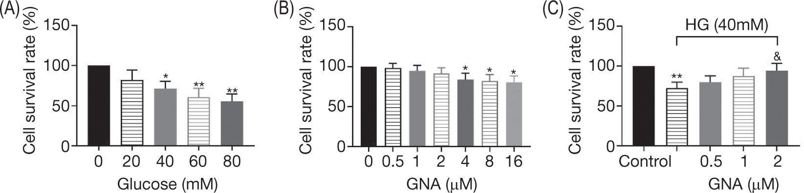
High concentrations of D-glucose (40, 60 or 80 μM) reduced the viability of HK-2 cells in a dose-dependent manner (Figure 1A), suggesting the cytoxicity of high glucose on renal tubular epithelial cells. Administration of gambogenic acid below 2-μM concentration had no significant effect on the viability of HK-2 cells (Figure 1B), while 4-μM concentration or more (4, 8 or 16 μM) decreased the viability of HK-2 cell line (Figure 1B). HK-2 cell line was co-incubated with 40-μM glucose and 0.5-, 1- or 2-μM gambogenic acid to investigate the nephroprotective effect of gambogenic acid against high glucose-caused decrease in cell viability. Indeed, the decreased viability of HK-2 cells caused by high glucose was increased by gambogenic acid in a dose-dependent manner (Figure 1C).
Figure 1. Gambogenic acid enhanced the viability of high glucose (HG)-treated HK-2 cells. (A) D-glucose reduced the viability of HK-2 cells in a dose-dependent manner. (B) Gambogenic acid at concentrations of 0, 0.5, 1 and 2 μM had no significant effect on the viability of HK-2 cells, while at concentrations of more than 4 μM (8 and 16 μM) decreased the viability of HK-2 cells. (C) Gambogenic acid enhanced the viability of high glucose-treated HK-2 cells in a dose-dependent manner. *, **vs. control, P < 0.05, P < 0.01. &vs. HG group, P < 0.05.
Gambogenic acid suppressed inflammation in high glucose-treated HK-2 cell line
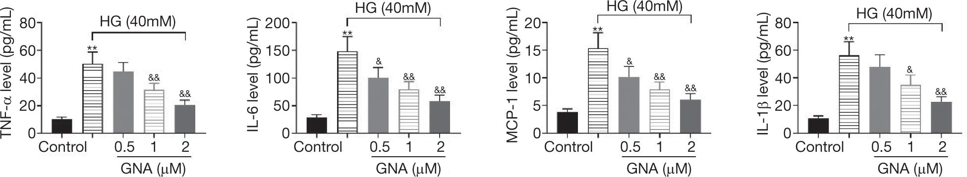
The levels of proinflammatory cytokines, TNF-α, IL-6, MCP-1 and IL-1β, were up-regulated in HK-2 cells after exposed to high glucose (40 μM) (Figure 2). However, gambogenic acid down-regulated the levels of TNF-α, IL-6, MCP-1 and IL-1β of high glucose-treated HK-2 cells (Figure 2) in a dose-dependent manner, indicating the anti-inflammatory effect of gambogenic acid on high glucose-treated HK-2 cells.
Figure 2. Gambogenic acid suppressed inflammation in high glucose (HG)-treated HK-2 cells. Gambogenic acid down-regulated the levels of TNF-α, IL-6, MCP-1, and IL-1β in high glucose-treated HK-2 cells. **vs. control, P < 0.01. &P < 0.05. &&vs. HG group, P < 0.01.
Gambogenic acid suppressed pyroptosis in high glucose-treated HK-2 cell line
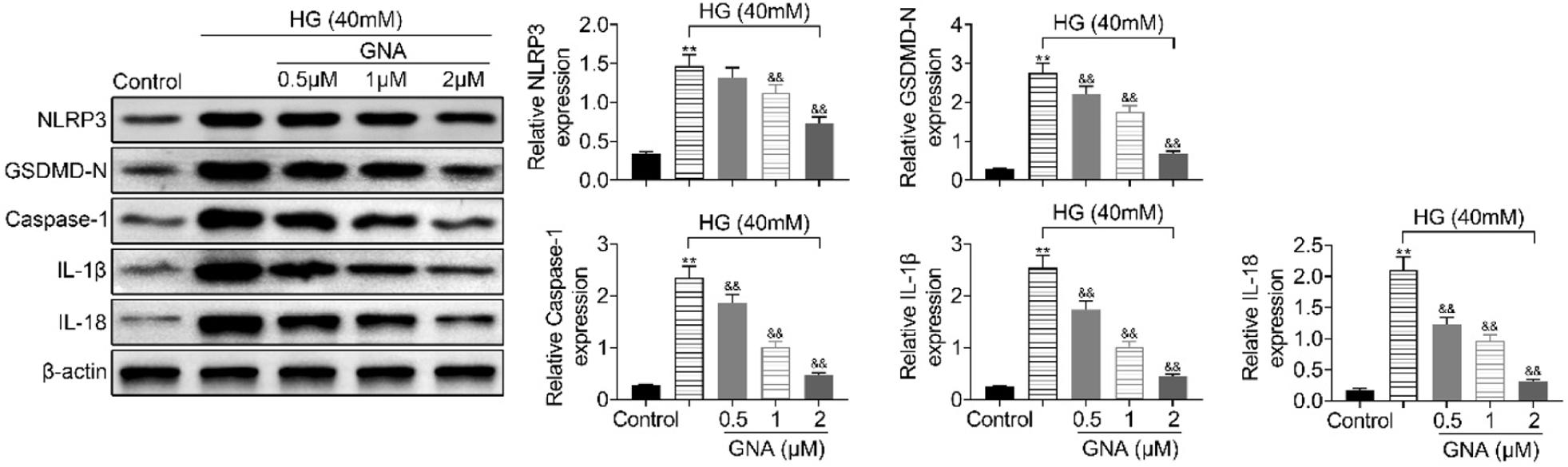
High glucose induces the up-regulation of NLRP3, GSDMD-N, caspase-1, IL-1β and IL-18 in HK-2 cells (Figure 3). However, the levels of NLRP3, GSDMD-N, caspase-1, IL-1β and IL-18 in high glucose-treated HK-2 cells are reduced by gambogenic acid in a dose-dependent manner (Figure 3). These results demonstrated that gambogenic acid suppresses pyroptosis in high glucose-treated HK-2 cell line.
Figure 3. Gambogenic acid suppressed pyroptosis in high glucose (HG)-treated HK-2 cells. Gambogenic acid down-regulated the levels of NLRP3, GSDMD-N, caspase-1, IL-1β and IL-18 in high glucose-treated HK-2 cells. **vs. control, P < 0.01. &&vs. HG group, P < 0.01.
Gambogenic acid suppressed the activation of AMPK–TXNIP signaling in high glucose-treated HK-2 cell line
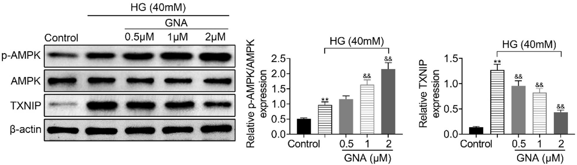
High glucose induces the up-regulation of TXNIP (Figure 4) and down-regulates the protein expression of p-AMPK in high glucose-treated HK-2 cells (Figure 4). Administration of gambogenic acid reduces the protein expression of TXNIP and promotes the protein expression of p-AMPK in high glucose-treated HK-2 cell line (Figure 4). Gambogenic acid suppresses the activation of AMPK–TXNIP signaling to protect HK-2 cell line against high glucose condition.
Figure 4. Gambogenic acid suppressed activation of AMPK–TXNIP signaling in high glucose (HG)-induced HK-2 cells. Gambogenic acid reduced the protein expression of TXNIP and promoted the protein expression of p-AMPK in high glucose-treated HK-2 cell line. **vs. control, P < 0.01. &&vs. HG group, P < 0.01.
Discussion
Chinese herbal medicines demonstrate long-term beneficial effects in the patients with diabetic nephropathy (Guo et al., 2021). For example, kolaviron, derived from Garcinia kola seeds, suppressed inflammation and oxidative stress in rats with diabetic nephropathy (Ayepola et al., 2014). Prenylated caged xanthones, derived from Garcinia hanburyi, are established to function as an inhibitor of protein tyrosine phosphatase 1B and thus involved in the progression and development of diabetes (Tan et al., 2017). Gambogic acid is a xanthonoid derived from Garcinia hanburyi that protects against high glucose- induced inflammatory response in diabetic retinopathy (Chen et al., 2021). The present study established that gambogic acid exerted protective effect against diabetic nephropathy by regulating cell viability, inflammation and pyroptosis.
Gambogic acid is regarded as a tumor suppressor through inhibiting the proliferation of tumor cells (Xu & Wantao, 2012). High concentration of gambogic acid (≥4 μM) in this study reduced the viability of HK-2 cells. However, its lower concentration (μ2 μM) increased the viability of high glucose-treated HK-2 cells. Hyperglycemia induces the inflammatory response of renal tubular epithelial cells, and contributes to the pathogenesis of diabetic nephropathy (Salti et al., 2020). Suppression of high glucose-induced inflammation relieves renal tubular injury in diabetic nephropathy (Su et al., 2020). Consistent with a previous study establishing that gambogenic acid inhibits lipopolysaccharide-induced up-regulation of IL-1α, IL-1β, TNF-α, IFN-β, IL-12β and IL-23α in macrophages (Akutsu et al., 2016), incubation with gambogenic acid down-regulates the levels of TNF-α, IL-6, MCP-1 and IL-1β in high glucose-treated HK-2 cells. Therefore, gambogenic acid protects renal tubular epithelial cells against high glucose condition through proliferative and anti-inflammatory effects. High glucose also induces the apoptosis of renal tubular epithelial cells through Rho kinase signal (Li et al., 2018), and hyperglycemia-induced apoptosis of renal tubular epithelial cells is the characteristic of diabetic nephropathy (Zhang et al., 2019). Gambogic acid exerts pro-apoptotic effect on tumor cells (Chen et al., 2015). The effect of gambogic acid on the apoptosis of high glucose-treated HK-2 cells must be investigated in the future research.
Pyroptosis, characterized by release of inflammatory factors (IL-1β and IL-18) through activation of caspase-1, is a pattern of programmed cell death and conceived as a recommendation for clinical diagnosis, targeted treatment and prognosis of kidney diseases (Zhang et al., 2021). Pyroptosis facilitates local inflammation and promotes the progression of diabetic complications (Mamun et al., 2021). Moreover, activation of NLRP3 inflammasome contributes to caspase-1-mediated pyroptosis and progression of diabetic nephropathy (Li et al., 2020). High glucose also induces the expression of protein levels of NLRP3, GSDMD, caspase-1, IL-1β and IL-18 in renal tubular epithelial cells, and suppression of NLRP3-mediated pyroptosis ameliorates high glucose-induced cell injury (Ke et al., 2020). A previous study has proved the suppressive effect of gambogic acid on NLRP3-mediated pyroptosis signaling in diabetic retinopathy (Chen et al., 2021). Results in this study demonstrated that gambogic acid reduced the levels of NLRP3, GSDMD, caspase-1, IL-1β and IL-18, thus exerting antidiabetic effect against high glucose.
AMPK signaling regulates cell growth and energy metabolism, thus implicating in the pathogenesis of diabetic retinopathy (Kubota et al., 2011). The level of p-AMPK was down-regulated in a mouse model with diabetic nephropathy, and activation of AMPK pathway promoted autophagy and reduced renal cell apoptosis, thus alleviating renal injury induced by high glucose (Song et al., 2021). AMPK promoted the activation of NLRP3 inflammasome to suppress hyperglycemia-induced pyroptosis in cardiomyocytes through degeneration of TXNIP (Wei et al., 2019). AMPK–TXNIP–NLRP3 signaling was involved in the pathogenesis of diabetic nephropathy. (An et al., 2020; Yang et al., 2021). Gambogic acid promoted the activation of AMPK pathway (Kim et al., 2014). Here, gambogic acid promoted the protein expression of p-AMPK and reduced the protein expression of TXNIP in high glucose-induced HK-2 cells, suggesting that AMPK–TXNIP was associated with gambogic acid-mediated diabetic retinopathy.
Conclusion
Gambogic acid exerts renal protective effects against high glucose condition by inhibiting inflammation and pyroptosis. Gambogic acid suppresses the activation of AMPK–TXNIP-mediated NLRP3 inflammasome to attenuate high glucose-induced injury in HK-2 cells. Therefore, gambogic acid could be a novel therapeutic avenue for diabetic nephropathy. However, the effect of gambogic acid on animal model with diabetic nephropathy must be investigated in the future research.
Competing interests
The authors state that there were no conflicts of interest to disclose.
Contribution of authors
Ningxu Li and Xiuying Wen designed and Mingjuan Tang conducted the experiments. Xiangmei Peng analyzed and interpreted the data. Qizhi Sheng and Ping Liu prepared the manuscript with contributions of all coauthors.
REFERENCES
Akutsu, H., Kaminishi, Y., Kurumisawa, S., Misawa, Y., 2016. Bioprosthetic aortic valve replacement 12 years after percutaneous aortic valvuloplasty in a young female adult with hope of pregnancy. Acute Medicine & Surgery 3(4): 364–368. 10.1002/ams2.178
An, X., Zhang, Y., Cao, Y., Chen, J., Qin, H. and Yang, L., 2020. Punicalagin protects diabetic nephropathy by inhibiting pyroptosis based on TXNIP/NLRP3 pathway. Nutrients 12: 1516. 10.3390/nu12051516
Ayepola, O.R., Cerf, M.E., Brooks, N.L. and Oguntibeju, O.O., 2014. Kolaviron, a biflavonoid complex of Garcinia kola seeds modulates apoptosis by suppressing oxidative stress and inflammation in diabetes-induced nephrotoxic rats. Phytomedicine 21: 1785–1793. 10.1016/j.phymed.2014.09.006
Chen, J., Li, L., Zhou, Y., Zhang, J. and Chen, L., 2021. Gambogic acid ameliorates high glucose-and palmitic acid-induced inflammatory response in ARPE-19 cells via activating Nrf2 signaling pathway: ex vivo. Cell Stress and Chaperones 26: 367–375. 10.1007/s12192-020-01182-1
Chen, J., Zhou, M., Zhang, Q., Xu, J. and Ouyang, J., 2015. Anticancer effect and apoptosis induction of gambogic acid in human leukemia cell line K562 in vitro. Medical Science Monitor: International Medical Journal of Experimental and Clinical Research 21: 1604–1610. 10.12659/MSM.893004
Ding, Z., Li, Y., Tang, Z., Song, X., Jing, F., Wu, H. and Lu, B., 2021. Role of gambogenic acid in regulating PI3K/Akt/NF-kβ signaling pathways in rat model of acute hepatotoxicity. Bioscience, Biotechnology, and Biochemistry 85: 520–527. 10.1093/bbb/zbaa039
Guo, J.C.-L., Pan, H.-C., Yeh, B.-Y., Lu, Y.C., Chen, J.-L., Yang, C.-W., Chen, Y.-C., Lin, Y.-H. and Chen, H.-Y., 2021. Associations between using chinese herbal medicine and long-term outcome among pre-dialysis diabetic nephropathy patients: a retrospective population-based cohort study. Frontiers in Pharmacology 12: 616522–616522. 10.3389/fphar.2021.616522
Hall, P.M., 2006. Prevention of progression in diabetic nephropathy. Diabetes Spectrum 19: 18. 10.2337/diaspect.19.1.18
He, Y., Ding, J., Lin, Y., Li, J., Shi, Y., Wang, J., Zhu, Y., Wang, K. and Hu, X., 2015. Gambogenic acid alters chemosensitivity of breast cancer cells to Adriamycin. BMC Complementary and Alternative Medicine 15: 181–181. 10.1186/s12906-015-0710-8
Ke, R., Wang, Y., Hong, S. and Xiao, L., 2020. Endoplasmic reticulum stress related factor IRE1α regulates TXNIP/NLRP3-mediated pyroptosis in diabetic nephropathy. Experimental Cell Research 396: 112293. 10.1016/j.yexcr.2020.112293
Keshvari, M., Rahmati, M., Mirnasouri, R. and Chehelcheraghi, F., 2020. Effects of endurance exercise and Urtica dioica on the functional, histological and molecular aspects of the hippocampus in STZ-Induced diabetic rats. Journal of Ethnopharmacology 256: 112801. 10.1016/j.jep.2020.112801
Kim, E., Kim, A., Kim, S., Chang, J., Ahn, C. and Chang, Y., 2014. Inhibition of mTORC1 induces loss of E-cadherin through AKT/GSK-3β signaling-mediated upregulation of E-cadherin repressor complexes in non-small cell lung cancer cells. Respiratory Research 15: 26. 10.1186/1465-9921-15-26
Kubota, S., Ozawa, Y., Kurihara, T., Sasaki, M., Yuki, K., Miyake, S., Noda, K., Ishida, S. and Tsubota, K., 2011. Roles of AMP-activated protein kinase in diabetes-induced retinal inflammation. Investigative Ophthalmology & Visual Science 52: 9142–9148. 10.1167/iovs.11-8041
Li, W.-N., Han, H., Jing, Z.-Y., Yang, X.-H., Zhang, Y. and Wei, J.-L., 2018. Mitochondrial oxidative damage and apoptosis induced by high glucose through Rho kinase signal pathway in renal tubular epithelial cells. Asian Pacific Journal of Tropical Medicine 11: 399–404. 10.4103/1995-7645.234769
Li, D.-X., Wang, C.-N., Wang, Y., Ye, C.-L., Jiang, L., Zhu, X. and Liu, Y.-J., 2020. NLRP3 inflammasome-dependent pyroptosis and apoptosis in hippocampus neurons mediates depressive-like behavior in diabetic mice. Behavioural Brain Research 391: 112684. 10.1016/j.bbr.2020.112684
Mamun, A.A., Wu, Y., Nasrin, F., Akter, A., Taniya, M.A., Munir, F., Jia, C. and Xiao, J., 2021. Role of pyroptosis in diabetes and its therapeutic implications. Journal of Inflammation Research 14: 2187–2206. 10.2147/JIR.S291453
Mei, W., Dong, C., Hui, C., Bin, L., Fenggen, Y., Jingjing, S., Cheng, P., Meiling, S., Yawen, H., Xiaoshan, W., Guanghui, W., Zhiwu, C. and Qinglin, L., 2014. Gambogenic acid kills lung cancer cells through aberrant autophagy. PloS One 9: e83604–e83604. 10.1371/journal.pone.0083604
Rahmati, M., Keshvari, M., Mirnasouri, R. and Chehelcheraghi, F., 2021. Exercise and urtica dioica extract ameliorate hippocampal insulin signaling, oxidative stress, neuroinflammation, and cognitive function in STZ-induced diabetic rats. Biomedicine & Pharmacotherapy 139: 111577. 10.1016/j.biopha.2021.111577
Rao, H., Jalali, J.A., Johnston, T.P. and Koulen, P., 2021. Emerging roles of dyslipidemia and hyperglycemia in diabetic retinopathy: molecular mechanisms and clinical perspectives. Frontiers in Endocrinology 12: 620045–620045. 10.3389/fendo.2021.620045
Rossing, P., Persson, F. and Frimodt-Møller, M., 2018. Prognosis and treatment of diabetic nephropathy: Recent advances and perspectives. Nephrologie & Therapeutique 14(Suppl 1): S31–S37. 10.1016/j.nephro.2018.02.007
Salti, T., Khazim, K., Haddad, R., Campisi-Pinto, S., Bar-Sela, G. and Cohen, I., 2020. Glucose induces IL-1α-dependent inflammation and extracellular matrix proteins expression and deposition in renal tubular epithelial cells in diabetic kidney disease. Frontiers in Immunology 11: 1270–1270. 10.3389/fimmu.2020.01270
Satirapoj, B. and Adler, S.G., 2015. Prevalence and management of diabetic nephropathy in western countries. Kidney Diseases (Basel, Switzerland) 1: 61–70. 10.1159/000382028
Song, S., Bao, S., Zhang, C., Zhang, J., Lv, J., Li, X., Chudhary, M., Ren, X. and Kong, L., 2021. Stimulation of AMPK prevents diabetes-induced photoreceptor cell degeneration. Oxidative Medicine and Cellular Longevity 2021: 5587340. 10.1155/2021/5587340
Su, J., Ren, J., Chen, H. and Liu, B., 2020. MicroRNA-140-5p ameliorates the high glucose-induced apoptosis and inflammation through suppressing TLR4/NF-κB signaling pathway in human renal tubular epithelial cells. Bioscience Reports 40: BSR20192384. 10.1042/BSR20192384
Tan, X.F., Uddin, Z., Park, C., Song, Y.H., Son, M., Lee, K.W. and Park, K.H., 2017. Competitive protein tyrosine phosphatase 1B (PTP1B) inhibitors, prenylated caged xanthones from Garcinia hanburyi and their inhibitory mechanism. Bioorganic & Medicinal Chemistry 25: 2498–2506. 10.1016/j.bmc.2017.03.010
Wei, H., Bu, R., Yang, Q., Jia, J., Li, T., Wang, Q. and Chen, Y., 2019. Exendin-4 protects against hyperglycemia-induced cardiomyocyte pyroptosis via the AMPK-TXNIP pathway. Journal of Diabetes Research 2019: 8905917–8905917. 10.1155/2019/8905917
Xu, W. and Wantao, C., 2012. Gambogic acid is a novel anticancer agent that inhibits cell proliferation, angiogenesis and metastasis. Anti-Cancer Agents in Medicinal Chemistry 12: 994–1000. 10.2174/187152012802650066
Yan, F., Wang, M., Chen, H., Su, J., Wang, X., Wang, F., Xia, L. and Li, Q., 2011. Gambogenic acid mediated apoptosis through the mitochondrial oxidative stress and inactivation of Akt signaling pathway in human nasopharyngeal carcinoma CNE-1 cells. European Journal of Pharmacology 652: 23–32. 10.1016/j.ejphar.2010.11.018
Yang, M., Luo, S., Jiang, N., Wang, X., Han, Y., Zhao, H., Xiong, X., Liu, Y., Zhao, C., Zhu, X. and Sun, L., 2021. DsbA-L ameliorates renal injury through the AMPK/NLRP3 inflammasome signaling pathway in diabetic nephropathy. Frontiers in Physiology 12: 659751–659751. 10.3389/fphys.2021.659751
Zhang, K.-J., Wu, Q., Jiang, S.-M., Ding, L., Liu, C.-X., Xu, M., Wang, Y., Zhou, Y. and Li, L., 2021. Pyroptosis: a new frontier in kidney diseases. Oxidative Medicine and Cellular Longevity 2021: 6686617–6686617. 10.1155/2021/6686617
Zhang, J., Zhao, X., Zhu, H., Wang, J., Ma, J. and Gu, M., 2019. Apigenin protects against renal tubular epithelial cell injury and oxidative stress by high glucose via regulation of NF-E2-related Factor 2 (Nrf2) pathway. Medical Science Monitor: International Medical Journal of Experimental and Clinical Research 25: 5280–5288. 10.12659/MSM.915038
Zhao, L., Zhang, H., Bao, J., Liu, J. and Ji, Z., 2015. Saikosaponin-d protects renal tubular epithelial cell against high glucose induced injury through modulation of SIRT3. International Journal of Clinical and Experimental Medicine 8: 6472–6481.
Zhao, Q., Zhong, J., Bi, Y., Liu, Y., Liu, Y., Guo, J., Pan, L., Tan, Y. and Yu, X., 2020. Gambogenic acid induces Noxa-mediated apoptosis in colorectal cancer through ROS-dependent activation of IRE1α/JNK. Phytomedicine 78: 153306.