
Download


Original Article
Genkwanin improves inflammatory injury in rats with septic lung injury by regulating NF-κB signaling pathway
Guixiang Qin1, Sunbang Yi2*
1Department of Emergency, Nantong First People’s Hospital, Nantong, Jiangsu Province, China;
2Department of Emergency, Wenzhou Central Hospital, Wenzhou, Zhejiang Province, China
Abstract
To assess possible effects of genkwanin (GKA) in septic lung injury and its related mechanisms. An animal model of cecal ligation and puncture (CLP)-induced acute lung injury was constructed. Histological analysis and wet–dry (W/D) ratio of the lung tissue were observed. The cell apoptosis in this model was analyzed by caspase activity detection, protein levels of B-cell lymphoma protein 2 (Bcl-2) and Bcl-2-associated X (Bax), and cell inflammation in CLP model; after GKA treatment, it was analyzed by enzyme-linked-immunosorbent serologic assay (ELISA). The involvement of nuclear factor kappa B (NF-κB) signaling pathway was evaluated by Immunoblot assay. We constructed an animal model of CLP-induced acute lung injury. Our data revealed that GKA reduced lung edema and inflammation in CLP mice. In addition, GKA reduced lung injury and apoptosis in CLP mice. Mechanically, our data in addition confirmed that GKA improved inflammatory injury in CLP mice by regulating NF-κB signaling pathway. Our data therefore confirmed that GKA could serve as a promising drug for the treatment of sepsis-induced acute lung injury.
Key words: sepsis, genkwanin (GKA), acute lung injury (ALI), CLP, apoptosis, NF-κB signaling pathway
*Corresponding Author: Sunbang Yi, Department of Emergency, Wenzhou Central Hospital, No. 252 Baili East Road, Wenzhou, Zhejiang Province 325000, China. Email: [email protected]
Received: 13 October 2021; Accepted: 3 November 2021; Published: 2 April 2022
© 2022 Codon Publications
This is an Open Access article distributed under the terms of the Creative Commons Attribution-NonCommercial-ShareAlike 4.0 International (CC BY-NC-SA 4.0). License (http://creativecommons.org/licenses/by-nc-sa/4.0/)
Introduction
Sepsis is one of the deadliest diseases worldwide and usually leads to multiple organ failure, primarily because of an uncontrollable inflammatory response (Kalantari and Rosner, 2021). However, the pathophysiological mechanisms associated with the development and treatment of sepsis remain unclear. Sepsis is usually associated with organ dysfunction caused by the patient’s weak defense against infection (Shimada et al., 2021). The lung is the most fragile and important organ in sepsis. Acute lung injury (ALI) is a common inflammatory disease induced by sepsis (Cokluk et al., 2021). Acute lung injury caused by sepsis has high morbidity and mortality (Yang et al., 2021). In order to improve the prognosis of patients, it is necessary to additionally study its pathogenesis and develop drugs that are more efficient.
Genkwanin (GKA) is one of the primary non-glycoylated flavonoids found in some herbal medicines, such as Daphne genkwa, rosemary, etc., with anti-inflammatory properties (Kawano et al., 1966). Genkwanin has a variety of pharmacological effects, including antibacterial, anti-plasmodium, and free radical scavenging, and can inhibit lipopolysaccharide (LPS)-induced inflammatory responses (Cottiglia et al., 2001; Kim et al., 2004; Kraft et al., 2003). Genkwanin has anti-rheumatoid arthritis effects in mice by inhibiting Janus kinase–signal transducer and activator of transcription (JAK–STAT) and nuclear factor kappa B (NF-κB) signaling pathways (Gao et al., 2014). In addition, GKA inhibits tumor cell proliferation by enhancing host immunity and lowering levels of inflammatory factors (Bao et al., 2019). Another study indicated that GKA suppressed 1-methyl-4-phenylpyridinium (MPP+)-induced cytotoxicity by inhibiting TLR4 inflammasome pathway in a cellular model of Parkinson’s disease (Ao et al., 2020). However, the role of GKA in septic lung injury and its related mechanisms remains unclear.
NF-κB plays a key role in inflammatory response because it induces secretion of pro-inflammatory cytokines such as Interleukin (IL)-6, IL-1β and tumor necrosis factor-α (TNF-α) as well as inducible Nitric Oxide Synthase (iNOS). In response to various stimuli (e.g., cytokines, DNA damage agents, and bacterial wall or viral proteins), inhibitor of nuclear factor kappa B (IκB) is dissociated and the activated transcription factors translocate to the nucleus, inducing a large number of target genes involved in cell growth, apoptosis, cell adhesion and inflammation. The NF-κB system is essential for regulating the innate immune response of host tissues.
In this study, we constructed an animal model of cecal ligation and puncture (CLP)-induced acute lung injury. Our data revealed that GKA could reduce lung edema and inflammatory injury, improve lung lesion, inhibit apoptosis and regulate NF-κB signaling pathway in septic lung injury mice. Our data therefore confirmed that GKA could serve as a promising drug for the treatment of sepsis-induced acute lung injury.
Materials and Methods
Sepsis lung injury model
Male Sprague Dawley (SD) mice (total number = 20, 5 per group) were bought from the Shanghai Laboratory Animal Center (SLAC; Shanghai, China) and maintained in a room with free access to normal chow diet and water in a 12-h light–dark cycle. All animal protocols were performed according to the Guide for the Care and Use of Laboratory Animals, 8th edition (National Institutes of Health, National Academies Press, US), and ethical approval was obtained from the Ethics Committee of Nantong First People’s Hospital. Animals were separated into four groups (five mice per group): (1) sham; (2) CLP group treated with vehicle; (3) GKA-treated groups were treated per os (through mouth) with GKA (5 and 10 mg/kg, respectively, suspended in 0.5% sodium carboxymethyl cellulose (CMC-Na) solution) every day. 5 mg/kg GKA-treated CLP group; and (4) 10 mg/kg GKA-treated CLP group. Then, the lower abdomen area of mice was shaved and sterilized after anesthetized with sodium pentobarbital. After incisions, the cecum was ligated below the ileocecal valve, followed by a single ‘through and through’ perforation (21-gauge needle). Incision was closed after the cecum was replaced in the abdomen.
Lung wet-to-dry (W/D) weight ratio measurement
The lung samples were dissected after the mice were sacrificed, and weighed immediately. Then the lung was dried until the weight was stable. The W/D weight ratio was obtained.
Cytokine measurement
The concentration of pro-inflammatory cytokines TNF-α, IL-1β, IL-10 and IL-6 was established in the blood and lung tissues with enzyme-linked immunosorbent assay (ELISA) kit according to manufacturer’s instructions (Kanglang, Shanghai, China).
hematoxylin & Eosin (H&E) staining
The lung tissue collected from all the groups were cut into slices. Slices were dehydrated through absolute alcohol and rehydrated. Slides were stained with hematoxylin for 4 min, rinsed, differentiated in 70% alcohol, stained in eosin Y, and cleared in xylenes before mounting. The hyperemia/congestion, edema and inflammation in the lung and alveolar collapse were accepted as lung injury. In order to examine the extent of lung injury, we considered the following five pathological features: (i) presence of exudate, (ii) hyperemia/congestion, (iii) intra-alveolar hemorrhage/detritus, (iv) cell infiltration and (v) cell proliferation. The severity parameters of each of these pathological features are as follows: 0 = nonpresence/absence, 1 = mild, 2 = moderate and, finally, 3 = severe injury. Scores are assessed with an overall score (0 to 15), and the sum of scores for different animals was averaged and plotted on a bar chart.
Cell apoptosis
For determining cell apoptosis, the activity of caspase-3 and caspase-9 was monitored with Caspase-3 kit and Caspase-9 kit, respectively. The activity of caspase-3 and caspase-9 was determined with the caspase-3 activity detection kit (ab252897; Abcam, Cambridge, UK) and caspase-9 activity detection kit (ab65607; Abcam), respectively, according to manufacturer’s instructions.
TUNEL assays were also used for the detection of apoptosis in lung tissues. Sliced sections were digested with 20-mg/mL proteinase K at 37°C for 15 min. Sections were then rinsed in phosphate-buffered saline (PBS) solution and added with 0.3% H2O2 for 10 min. The sections were incubated with 0.1% sodium citrate and 0.1% Triton X-100 solution for 2 min. Then TUNEL reaction mixture with terminal deoxynucleotidyltransferase (TdT) (Sigma-Aldrich, St. Louis, MO, USA) was utilized for sections at 37°C under humidified conditions followed by DAPI (4',6-diamidino-2-phenylindole) staining. Each image was captured using a confocal microscope.
Immunoblot assay
Nuclear protein NF-κB and cytosolic IκBα were measured from isolated nuclear and cytosol proteins through nuclear extraction kit (ab113474). Proteins were extracted with radioimmunoprecipitation assay (RIPA) buffer (Cell Signaling). Then the cell samples were collected and subjected to 10% sodium dodecyl sulfate–polyacrylamide gel electrophoresis (SDS-PAGE), and transferred onto polyvinylidene difluoride (PVDF) membranes, followed by blocking with 5% bovine serum albumin (BSA) in TBST (Tris-Buffered Saline+Tween 20) buffer. Subsequently, membranes were conjugated with primary antibodies targeting Bcl-2-associated X (Bax, 1:1,000; Abcam, Cambridge, UK), B-cell lymphoma protein 2 (Bcl-2, 1:1,000; Abcam), anti-p65 (1:2,000, Abcam), anti-p-p65 (1:1,000, Abcam), anti-p-IκBα (1:1,000, Abcam), anti-IκBα (1:1,000, Abcam) and glyceraldehyde 3-phosphate dehydrogenase (GAPDH, 1:1,0000; Abcam) for 2 h at room temperature. Subsequently the membranes were incubated with specific secondary antibodies at room temperature for 1 h. The blots were analyzed with ECL kit.
Statistical Analysis
Data were displayed as mean ± SD. Statistical analysis was performed using GraphPad. Significance was assessed by analysis of variance (ANOVA). P < 0.05 was considered as level of significance.
Results
GKA alleviates CLP-induced lung edema and inflammation
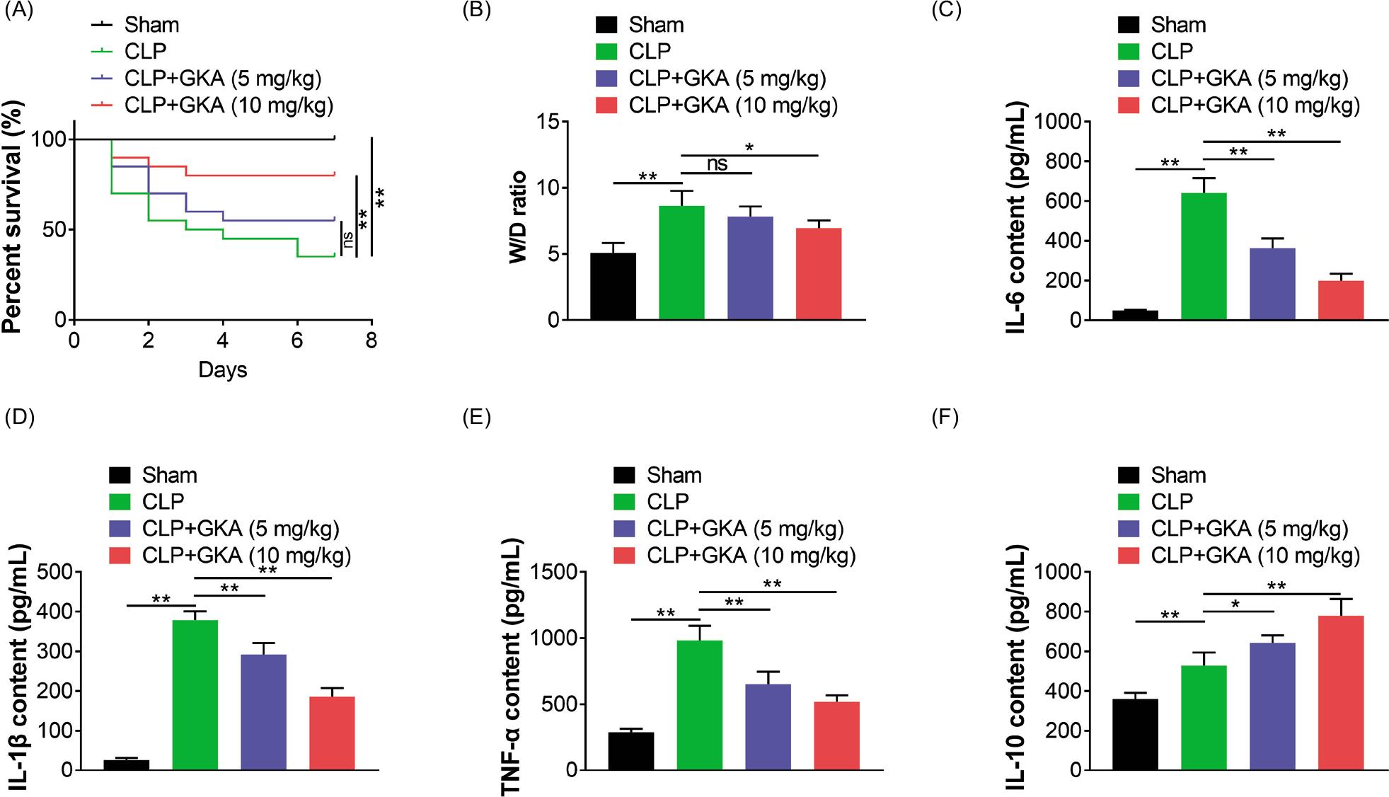
In order to explore the therapeutic effect of GKA on CLP-induced lung injury in mice, we first constructed four groups of mice with and without GKA pretreatment. After construction of CLP model, the W/D ratio of the lung tissue was assessed. CLP induced the increased ratio of W/D in mice. GKA treatment reversed the elevated W/D ratio induced by CLP (Figure 1A). In order to delineate the role of GKA on sepsis-induced inflammatory cytokine production, concentration of TNF-α, IL-6, IL-10 and IL-1β in the blood of mice was determined. As shown in Figures 1B–1E, sepsis stimulation significantly induced elevated levels of TNF-α, IL-6, IL-10 and IL-1β in mice. However, administration of GKA relieved the increase of these pro-inflammatory cytokines in the blood of mice. IL-10 was further increased by GKA treatment (Figure 1E). Furthermore, it was noticed that GKA alleviated CLP-induced lung inflammation in the lung tissues of mice, with the decreased level of TNF-α, IL-6, IL-10 and IL-1β (Figure 2A). Therefore, we believed that GKA alleviated CLP-induced lung edema and inflammation.
Figure 1. GKA alleviates CLP-induced lung edema and inflammation. (A) The survival rate of mice in sham, CLP, CLP + 5 mg/kg GKA, and CLP+10 mg/kg GKA groups (20 in each group). (B) The W/D ratio of lung in sham, CLP, CLP + 5 mg/kg GKA, and CLP + 10 mg/kg GKA groups. (C)–(F) The IL-6, IL-10, IL-1β, and TNF-α levels in the blood of sham, CLP, CLP + 5 mg/kg GKA, and CLP + 10 mg/kg GKA groups. *P < 0.05, **P< 0.01.
Figure 2. GKA suppresses CLP-induced cell inflammation and apoptosis in mice with lung injury. (A) The IL-6, IL-10, IL-1β, and TNF-α levels in the lung tissues of sham, CLP, CLP + 5 mg/kg GKA, and CLP+10 mg/kg GKA groups. (B) TUNEL assays demonstrated the cell apoptosis levels of lung tissues in sham, CLP, CLP + 5 mg/kg GKA, and CLP + 10 mg/kg GKA groups. **P < 0.01.
GKA relieves CLP-induced lung injury in mice
Histological changes in all groups were analyzed through H&E staining. CLP group revealed obvious lung hyperemia/congestion and alveolar collapse compared with the sham group (Figure 3A). GKA treatment alleviated phenomenon of lung edema and inflammatory cell infiltration in a dose-dependent manner (Figure 3B). Moreover, the increased lung injury score induced by CLP model was reduced in GKA treated mice. Taken together, these results indicated that GKA could attenuate CLP-induced lung injury.
Figure 3. GKA relieves CLP-induced lung injury in mice. (A) Histological changes in lung tissue in sham, CLP, CLP + 5 mg/kg GKA, and CLP + 10 mg/kg GKA groups. (B) The lung injury score in sham, CLP, CLP + 5 mg/kg GKA, and CLP + 10 mg/kg GKA groups. *P < 0.05, **P < 0.01.
GKA suppresses CLP-induced cell apoptosis in mice with lung injury
The effect of GKA on cell apoptosis was examined by the activity of caspase-3 and caspase-9. Cell apoptosis increased in the CLP group as revealed by elevated caspase-3 and caspase-9 activity. GKA treatment reduced cell apoptosis (Figures 4A and 4B). In addition, cell apoptosis was induced in the CLP group as revealed by elevated Bax and reduced Bcl-2 levels. GKA relieved Bax level and enhanced Bcl-2 protein level (Figure 4C). Our data further confirmed through TUNEL assays that GKA suppressed cell apoptosis in lung tissues (Figure 2B). These data suggested that GKA treatment exerted reduced cell apoptosis in the lung injury of mice.
Figure 4. GKA suppresses CLP-induced cell apoptosis in mice with lung injury. (A) and (B) Activity of caspase-3 and caspase-9 in sham, CLP, CLP + 5 mg/kg GKA, and CLP + 10 mg/kg GKA groups. (C) Protein levels of Bax and Bcl-2 in four groups were detected. *P < 0.05, **P < 0.01.
GKA represses CLP injury via NF-κB signaling pathway
In order to reveal potential mechanism, the activation of NF-κB signaling pathway was analyzed (Figure 5A). We also observed the enhanced p-p65, p-IκBα and reduced IκBα in CLP-treated cells (Figure 5A). GKA treatment partially relieved these altered proteins in a dose-dependent manner, further implying that GKA alleviates CLP-induced lung injury by regulating NF-κB signaling pathway.
Figure 5. GKA represses CLP injury via NF-κB signaling pathway. Immunoblot assay detected the protein level of p-p65 and p-IkBa in sham, CLP, CLP+5 mg/kg GKA, and CLP+10 mg/kg GKA groups. *P < 0.05, **P < 0.01.
Discussion
Sepsis is one of the leading causes of death in critically ill patients. It infects millions of people worldwide annually, kills a quarter or more of them, and the incidence is still rising (Cokluk et al., 2021). Sepsis is also a common cause of acute lung injury and acute respiratory distress syndrome, which seriously affects the therapeutic results of the disease (Yang et al., 2021). Currently, there are two primary mechanisms of acute lung injury induced by sepsis (van Bockxmeer et al., 2021). The first one is believed to be caused by the entry of activated neutrophils into the lung, and the other is believed to be caused by inflammation, apoptosis and dysfunction of pulmonary endothelial cells after activation, thus leading to lung injury (Ren et al., 2021). The most commonly used model to study acute lung injury is CLP, which can produce septic-induced acute lung injury. In this study, we successfully constructed a CLP-induced acute lung injury model. Our data further confirmed that GKA improved inflammatory injury in CLP mice with acute lung injury. We therefore thought that GKA could serve as a promising drug for the treatment of sepsis-induced acute lung injury.
Performing H&E staining and ELISA, we found that GKA reduced lung edema and inflammation in CLP mice. Further, through ELISA and Immunoblot assay, our data confirmed that GKA stimulated apoptosis in CLP mice. These findings confirmed the effects of GKA on acute lung injury of CLP mice. In fact, the multiple biological activities of GKA have been widely established (Bao et al., 2019). Genkwanin suppressed MPP+-induced cytotoxicity by inhibiting TLR4 inflammasome pathway in Parkinson’s disease (Jiang et al., 2014; Li et al., 2017). In addition, GKA enhanced antitumor efficacy against breast cancer (Wang et al., 2015). Genkwanin ameliorated adjuvant-induced arthritis in mice by suppressing NF-κB signaling pathway (Yuan and Wang, 1995). Similarly, we also found that GKA improved inflammatory injury in CLP mice by regulating NF-κB signaling pathway. Taken together, these studies demonstrated that GKA had a variety of pharmacological effects, and could serve as a drug for various diseases.
In this study, we also revealed that GKA ameliorated acute lung injury in CLP mice by suppressing NF-κB signaling pathway (Liu et al., 2011). NF-κB plays a key role in inflammatory response because of the induction of the secretion of pro-inflammatory cytokines (Zhou et al., 2018). Importantly, the NF-κB system is essential for regulating the innate immune response of host tissues. In addition, this pathway is widely involved in the pathology of sepsis and acute lung injury (Zhang et al., 2021). Several promising drugs could be used for the treatment of acute lung injury (Yu et al., 2014). For example, Berberine attenuated sepsis by suppressing FOXA1 expression and NF-κB signaling pathway by the induction of miR-132-3p (Yang et al., 2017). Gadolinium chloride reduced inflammatory response and preserved intestinal barrier function in sepsis mice through this pathway. In addition, dehydrocorydaline protected against sepsis-induced myocardial injury by regulating the NF-κB signaling pathway (Wang et al., 2021). As a comparison, Astragaloside IV could attenuate polymicrobial sepsis-induced cardiac dysfunction through NF-κB signaling pathway in mice (Wakana et al., 2008). These studies, together with our findings, could act as a promising target for the treatment of sepsis.
Conclusion
We constructed an animal model of CLP-induced acute lung injury. Our data revealed that GKA could reduce lung edema and inflammatory injury, inhibit apoptosis and mediate NF-κB signaling pathway in septic acute lung injury mice. Therefore, our data confirmed that GKA could serve as a promising drug for the treatment of sepsis-induced acute lung injury.
Competing Interests
The authors state that there are no conflicts of interest to disclose.
Contribution of Authors
Guixiang Qin and Sunbang Yi designed and conducted the study. Both authors supervised data collection, and analyzed and interpreted the data. They prepared, reviewed, read, and approved the final draft of the manuscript for publication
REFERENCES
Ao, H., Li, Y., Li, H., Wang, Y., Han, M., Guo, Y., Shi, R., Yue, F. and Wang, X., 2020. Preparation of hydroxy genkwanin nanosuspensions and their enhanced antitumor efficacy against breast cancer. Drug Delivery 27: 816–824. 10.1080/10717544.2020.1770372
Bao, Y., Sun, Y.W., Ji, J., Gan, L., Zhang, C.F., Wang, C.Z. and Yuan, C.S., 2019. Genkwanin ameliorates adjuvant-induced arthritis in rats through inhibiting JAK/STAT and NF-kappaB signaling pathways. Phytomedicine 63: 153036. 10.1016/j.phymed.2019.153036
Cokluk, E., Doganay, S., Ramazan Sekeroglu, M., Betul Tuncer, F., Cakiroglu, H. and Boz, M., 2021. Investigation of the effect of melatonin administration on inflammatory mediators; MMP-2, TGF-beta and VEGF levels in rats with sepsis. International Journal of Clinical Practice e14832. 10.1111/ijcp.14832
Cottiglia, F., Loy, G., Garau, D., Floris, C., Casu, M., Pompei, R. and Bonsignore, L., 2001. Antimicrobial evaluation of coumarins and flavonoids from the stems of Daphne gnidium L. Phytomedicine 8: 302–305. 10.1078/0944-7113-00036
Gao, Y., Liu, F., Fang, L., Cai, R., Zong, C. and Qi, Y., 2014. Genkwanin inhibits proinflammatory mediators mainly through the regulation of miR-101/MKP-1/MAPK pathway in LPS-activated macrophages. PLoS One. 9: e96741. 10.1371/journal.pone.0096741
Jiang, C.P., He, X., Yang, X.L., Zhang, S.L., Li, H., Song, Z.J., Zhang, C.F., Yang, Z.L. and Li, P., 2014. Intestinal absorptive transport of Genkwanin from Flos genkwa using a single-pass intestinal perfusion rat model. American Journal of Chinese Medicine 42: 349–359. 10.1142/S0192415X14500232
Kalantari, K. and Rosner, M.H., 2021. Recent advances in the pharmacological management of sepsis-associated acute kidney injury. Expert Review of Clinical Pharmacology 1–11. 10.1080/17512433.2021.1978287
Kawano, N., Miura, H. and Matsuishi, E., 1966. The partial demethylation of flavones. I. Preparation of genkwanin. Chemical and Pharmaceutical Bulletin (Tokyo) 14: 299–300. 10.1248/cpb.14.299
Kim, A.R., Zou, Y.N., Park, T.H., Shim, K.H., Kim, M.S., Kim, N.D., Kim, J.D., Bae, S.J., Choi, J.S. and Chung, H.Y., 2004. Active components from Artemisia iwayomogi displaying ONOO(-) scavenging activity. Phytotherapy Research 18: 1–7. 10.1002/ptr.1358
Kraft, C., Jenett-Siems, K., Siems, K., Jakupovic, J., Mavi, S., Bienzle, U. and Eich, E., 2003. In vitro antiplasmodial evaluation of medicinal plants from Zimbabwe. Phytotherapy Research 17: 123–128. 10.1002/ptr.1066
Li, Y., Hong, J., Li, H., Qi, X., Guo, Y., Han, M. and Wang, X., 2017. Genkwanin nanosuspensions: a novel and potential antitumor drug in breast carcinoma therapy. Drug Delivery 24: 1491–1500. 10.1080/10717544.2017.1384519
Liu, J.L., Ma, H.P., Lu, X.L., Sun, S.H., Guo, X. and Li, F.C., 2011. NF-kappa B induces abnormal centrosome amplification by upregulation of CDK2 in laryngeal squamous cell cancer. International Journal of Oncology 39: 915–924. 10.3892/ijo.2011.1125
Ren, C., Yao, R.Q., Wang, L.X., Li, J.C., Chen, K.W., Wu, Y., Dong, N., Feng, Y.W. and Yao, Y.M., 2021. Antagonism of cerebral high mobility group box 1 ameliorates dendritic cell dysfunction in sepsis. Frontiers in Pharmacology 12: 665579. 10.3389/fphar.2021.665579
Shimada, B.K., Boyman, L., Huang, W., Zhu, J., Yang, Y., Chen, F., Kane, M.A., Yadava, N., Zou, L., Lederer, W.J., Polster, B.M. and Chao, W., 2021. Pyruvate-driven oxidative phosphorylation is downregulated in sepsis-induced cardiomyopathy: a study of mitochondrial proteome. Shock. 10.1097/SHK.0000000000001858
van Bockxmeer J.J., Shetty A., Robertson T. and Thomas Y., 2021. Closing the sepsis gap: an exploration of sepsis presentations at a remote north Australian emergency department. Rural Remote Health 21: 5979. 10.22605/RRH5979
Wakana, Y., Takai, S., Nakajima, K., Tani, K., Yamamoto, A., Watson, P., Stephens, D.J., Hauri, H.P. and Tagaya, M., 2008. Bap31 is an itinerant protein that moves between the peripheral endoplasmic reticulum (ER) and a juxtanuclear compartment related to ER-associated Degradation. Molecular Biology of the Cell 19: 1825–1836. 10.1091/mbc.E07-08-0781
Wang, H., Huang, X., Xu, P., Liu, X., Zhou, Z., Wang, F., Li, J., Wang, Y., Xian, X., Liu, G. and Huang, W., 2021. Apolipoprotein C3 aggravates diabetic nephropathy in type 1 diabetes by activating the renal TLR2/NF-kappaB pathway. Metabolism 154740. 10.1016/j.metabol.2021.154740
Wang, X., Song, Z.J., He, X., Zhang, R.Q., Zhang, C.F., Li, F., Wang, C.Z. and Yuan, C.S., 2015. Antitumor and immunomodulatory activity of genkwanin on colorectal cancer in the APC(Min/+) mice. International Immunopharmacology 29: 701–707. 10.1016/j.intimp.2015.09.006
Yang, J., Miao, X., Guan, Y., Chen, C., Chen, S., Zhang, X., Xiao, X., Zhang, Z., Xia, Z., Yin, T., Hei, Z. and Yao, W., 2021. Microbubble functionalization with platelet membrane enables targeting and early detection of sepsis-induced acute kidney injury. Advanced Healthcare Materials e2101628. 10.1002/adhm.202101628
Yang, K., Li, W.F., Yu, J.F., Yi, C. and Huang, W.F., 2017. Diosmetin protects against ischemia/reperfusion-induced acute kidney injury in mice. Journal of Surgical Research 214: 69–78. 10.1016/j.jss.2017.02.067
Yu, G., Wan, R., Yin, G., Xiong, J., Hu, Y., Xing, M., Cang, X., Fan, Y., Xiao, W., Qiu, L., Wang, X. and Hu, G., 2014. Diosmetin ameliorates the severity of cerulein-induced acute pancreatitis in mice by inhibiting the activation of the nuclear factor-kappa B. International Journal of Clinical and Experimental Pathology 7: 2133–2142.
Yuan, S. and Wang, Z., 1995. Influence of processing on contents of genkwanin in flos Genkwa. Zhongguo Zhong Yao Za Zhi 20: 340–342, 382.
Zhang, Q., Xue, T., Guan, J., Wang, W., Shi, J., Lu, J. and Jiang, X., 2021. Irigenin alleviates angiotensin II-induced oxidative stress and apoptosis in HUVEC cells by activating Nrf2 pathway. Drug Development Research 10.1002/ddr.21802
Zhou, H., Wang, J., Zhu, P., Zhu, H., Toan, S., Hu, S., Ren, J. and Chen, Y., 2018. NR4A1 aggravates the cardiac microvascular ischemia reperfusion injury through suppressing FUNDC1-mediated mitophagy and promoting Mff-required mitochondrial fission by CK2alpha. Basic Research in Cardiology 113: 23. 10.1007/s00395-018-0682-1