
Download
Original Article
Chemical properties of Indonesian aloe vera and its hepatoprotective activity by targeting reactive oxygen species and TACE protein: comparison between gel and whole parts
Sari Haryanti1, Mujiyanto Mujiyanto2, Nur Maulidah Rahmah3, Ade Erma Suryani4, Khoirun Nisa4, Santosh Chokkakula5, Soon Woong Chang6, Balasubramani Ravindran6,7*, Ravishankar Ram Mani8,9*, Sri Handayani4*
1Research Center for Pharmaceutical Ingredients and Traditional Medicine, National Research and Innovation Agency Republic of Indonesia (BRIN), Cibinong Science Center, Jalan Raya Jakarta-Bogor, West Java Indonesia;
2Research Center for Public Health and Nutrition, National Research and Innovation Agency Republic of Indonesia (BRIN), Cibinong Science Center, Jalan Raya Jakarta-Bogor, West Java, Indonesia;
3Master Program of Biotechnology, The Graduate School of Universitas Gadjah Mada, Jl. Teknika Utara, Yogyakarta, Indonesia;
4Research Center for Food Technology and Processing, National Research and Innovation Agency Republic of Indonesia (BRIN), JL. Jogja-Wonosari, Yogyakarta, Indonesia;
5Department of Microbiology, Chungbuk National University Medical College, Cheongju, South Korea;
6Department of Civil & Energy System Engineering, Kyonggi University, Suwon, Gyeonggi-Do, 16227, South Korea;
7Department of Microbiology, Faculty of Arts Science Commerce and Management, Karpagam Academy of Higher Education, Coimbatore, Tamil Nadu, India;
8Department of Pharmaceutical Biology, Faculty of Pharmaceutical Sciences, UCSI University, Cheras, 56000, Kuala Lumpur, Malaysia;
9Cellular and Molecular Biochemistry, Research and Development Wing, Sree Balaji Medical College and Hospital, Bharath Institute of Higher Education and Research (BIHER), Chromepet, Chennai, 600044, Tamil Nadu, India
Abstract
This study evaluated the chemical properties of Indonesian aloe vera and compared the hepatoprotective activity of aloe gel (AO) and whole plant (WAO) against H2O2-induced toxicity in HepG2 cells. Chemical characterization was performed using ultraviolet-visible (UV-Vis), Fourier transform infrared (FTIR) spectroscopy, liquid chromatography–high-resolution mass spectrometry (LC-HRMS), and high-performance liquid chromatography analyses, and compound interactions with tumor necrosis factor (TNF)-α converting enzyme (TACE/ADAM17) were examined. AO contained a higher level of O-acetyl polysaccharides (10.36 ± 0.63% by mass [b/b]) than WAO (6.39 ± 0.16% b/b). Aloin A was rich in WAO (1,342.3 ± 23.94 ppm b/b) but absent in AO. FTIR spectra were similar for both samples, while LC-HRMS profiling revealed phenolic, flavonoid, and terpenoid compounds in AO and WAO. Various concentrations of AO effectively inhibited H2O2-induced toxicity in HepG2 cells, with 200 μg/mL showing the highest protective effect (22.44 ± 6.04%). In contrast, WAO reduced toxicity at lower concentrations but became cytotoxic at higher concentrations. Both extracts decreased intracellular reactive oxygen species (ROS) compared to H2O2 alone. At 200 μg/mL, AO and WAO reduced ROS level, compared to H2O2 alone, by 0.964±0.241 fold and 0.768±0.128 fold, respectively (P < 0.05). Docking studies showed squalene (C30H50) from AO exhibited the strongest affinity for TACE protein with a docking score of -1,16.6±2.54. Overall, AO extract may serve as a potent hepatoprotective agent.
Key words: aloe vera, aloe gel, hepatoprotective, TNF-α converting enzyme (TACE), squalene
*Corresponding Authors: Balasubramani Ravindran, Department of Civil & Energy System Engineering, Kyonggi University, Suwon, Gyeonggi-Do, 16227, South Korea; Department of Microbiology, Faculty of Arts Science Commerce and Management, Karpagam Academy of Higher Education, Coimbatore, Tamil Nadu, India. Emails: [email protected]; [email protected]; Ravishankar Ram Mani, Department of Pharmaceutical Biology, Faculty of Pharmaceutical Sciences, UCSI University, Cheras 56000, Kuala Lumpur, Malaysia. Email: [email protected]; Sri Handayani, Research Center for Food Technology and Processing, National Research and Innovation Agency Republic of Indonesia (BRIN), JL. Jogja - Wonosari, km 31.5, Yogyakarta, Indonesia. Email: [email protected]
Academic Editor: Gulden Goksen, Tarsus University, Istanbul, Turkey
Received: 19 March 2025; Accepted: 1 September 2025; Published: 29 December 2025
© 2025 Codon Publications
This is an Open Access article distributed under the terms of the Creative Commons Attribution-NonCommercial-ShareAlike 4.0 International (CC BY-NC-SA 4.0). License (http://creativecommons.org/licenses/by-nc-sa/4.0/)
Introduction
Aloe vera is a native plant of southern and eastern Africa (Adlakha et al., 2022). Nevertheless, it is easily cultivated globally, including in several regions of Indonesia. Different environments affect the plant metabolite composition (Quispe et al., 2018). Aloe vera is a spiky cactus-like xerophyte belonging to the Liliaceae family. As a plant having a high content of water, aloe vera contains 0.5–1.0% of active compounds. Several polysaccharides, such as acemannan, arabinan, galactomannan, and glucomannan, have been identified in aloe vera gel (AO). Acemannan, the major polysaccharide in AO, ameliorates wound-healing of fibroblast cells. Minor amounts of various sterol, phenol, and flavonoid compounds, including kaempferol, naringenin, quercetin, and apigenin and its derivatives, are also determined in AO (Handayani et al., 2021). Several foods, beverages, and cosmetics use aloe plants as their ingredient because of its bioactive properties that benefit our health (Chandra et al., 2024). Polysaccharides and phenolic and flavonoid compounds contained in aloe plant have nutraceutical effects. These active compounds have prevention and protection attributes for inflammation, wounds, aging, and cancer proliferation as well as toward overproduction of free radicals (Añibarro-Ortega et al., 2019; Babu and Noor, 2021; Handayani et al., 2021, 2023; Quispe et al., 2018).
Aloe vera is commonly used as a topical remedy to treat sunburns and heal wounds. Promisingly, aloe beverage also provides many health benefits, such as reduce cholesterol and triglycerides, mitigate oxidative stress, fortify the immune system, alleviate nausea, ameliorate adverse effects of radiation therapy, promote hair growth, diminish arthritis, relieve joint and muscle discomfort, lower blood glucose levels, improve digestive functioning, alleviate acid reflux symptoms, enhance blood oxygenation, address dental and gum diseases, and decelerate the aging process (Akbari et al., 2022; Añibarro-Ortega et al., 2019; Guo and Mei, 2016). Furthermore, promotion of healthier aloe-based beverages enhances the nation’s socioeconomic status while also serving as a hepatoprotective agent. In Indonesia, despite numerous aloe vera products being produced, limited research is focused on the processing of aloe beverages, resulting in a lack of information on distinct aloin contained in AO and whole aloe plant (WAO), related to its toxicity level.
Aloe vera, particularly WAO leaves, contains high levels of aloin. Aloin is a major anthraquinone discovered in aloe perennial. Aloin has anticancer activity through the induction of cell death or apoptosis (Hu et al., 2022). However, several studies reported that aloin also has toxicity and carcinogenic effects depending on its concentration (Guo and Mei, 2016; Yang et al., 2022). The usage of aloe plants as a functional food ingredient requires a more comprehensive toxicity analysis, so that safe functional foods are produced. The EFSA Panel on Food Additives and Nutrient Sources Added to Food (ANS) and the US Food and Drug Administration (FDA) have set up 1.2–2.4 mg as maximum concentration of aloin for daily oral administration. The International Aloe Science Council (IASC) and the European Economic Community (EEC) established a quality standard of not more than 10 ppm (10 mg/kg) for aloin A and aloin B in all aloe vera products meant for oral consumption (EFSA Panel on Food Additives and Nutrient Sources added to Food (ANS) et al., 2018).
The liver is an important organ for detoxification of toxic substances. Therefore, this causes high potential damage to the liver. The mechanism of cell toxicity commonly involves reactive oxygen species (ROS), tumor necrosis factor (TNF)-α, and TNF-α converting enzyme (TACE). In normal conditions, the production of highly reactive ROS is an important feature of cellular systems, including fertilization, ovulation, arachidonic acid metabolism, and phagocytosis. However, the multiple times higher production of ROS indicates pathological complications (Chaudhary et al., 2023). The production of ROS in the cells shows the level of oxidative stress (Handayani et al., 2021). The liver is the main target of ROS under oxidative stress. Toxic substances induce hepatotoxicity by creating free radicals, including ROS (Handayani et al., 2021). On the other hand, as a response to oxidative stress, cytokines, such as TNF-α, are released and trigger apoptosis and inflammation (Chaudhary et al., 2023). Meanwhile, the over-expression of TNF-α in several inflammation cases was caused by the increasing levels of TNF-α converting enzyme (Lakshmi et al., 2019). As the involvement of TNF-α and its converting enzyme is increasingly recognized in liver inflammation, natural agents such as aloe vera have gained interest for their potential to interfere in these pathways and offer hepatoprotective benefits.
Hepatoprotective agents prevent toxic compounds causing liver damage and protect the liver from diseases. Compounds that have anti-inflammation, antioxidant, and inhibition of cell death have a role in hepatoprotective activity. The synergistic hepatoprotective effect may be driven by several phytochemicals discovered in AO (Handayani et al., 2021). Various studies revealed the protective effect of aloe vera on mice liver (Gupta et al., 2019, 2023; Padmanabhan and Jangle, 2014). Active phytochemicals in aloe vera reveal their hepatoprotective effects through various mechanisms, including protection against ROS and TNF-α (Handayani et al., 2021).
The in silico studies revealed the affinity of several compounds detected in aloe vera toward TNF-α receptor (Chinnadurai et al., 2024; Kaloni et al., 2019). Nevertheless, the in silico study on the affinity of different compounds detected in aloe vera toward the TACE enzyme is still limited. While numerous studies have demonstrated the general hepatoprotective effects of aloe vera, most have not distinguished between AO and WAO components related to specific molecular pathways. Moreover, limited research has addressed the role of aloe vera phytochemicals in modulating ROS and TACE activity under oxidative stress conditions. Zhou and Zhao (2021) stated in their study that H2O2 is one type of free radical production inducer that causes cell toxicity. Most of the superoxide anions produced by mitochondria are mutated into H2O2 (Chaudhary et al., 2023). In addition, the HepG2 cell is used as a model for liver injuries, including hepatoprotective activity (Al-Sheddi et al., 2024; Lee et al., 2021; Sahoo et al., 2023). Using this model, the present work aims to study the hepatoprotective activity of AO and WAO extract in H2O2-induced HepG2 cells, particularly in relation to ROS modulation and TACE enzyme involvement. To support this investigation, a foundational chemical characterization of aloe vera extracts was conducted to obtain prominent metabolites while minimizing aloin content. This was intended to determine the effective and safest functioning of aloe vera for potential therapeutic application.
Materials and Methods
Sample preparation
The aloe vera plant used in this study was obtained from the local plantation in Gunungkidul Region of Indonesia. This plant was authenticated as Aloe vera (L.) Burm.f. by Medicinal Plant and Traditional Medicine Research and Development Centre, Ministry of Health, Republic of Indonesia (No. KM.04.01/2/162/2021). WAO and AO from aloe leaves were sliced and dried at 50°C using an oven (Memmert, Germany). A part of AO was crushed using a blender to make AO juice (AJ). Then, WAO, AO, and AJ were separately extracted using 70% ethanol (Merck, Germany) to prepare ethanol extracts.
Soil analysis
The aloe plantation soil was collected from three locations. The soil from the same area that had not planted aloe vera was used as a control. Soil texture was analyzed at the Faculty of Geography, Universitas Gadjah Mada (No. 2/GLMB/V/2023). The soil components were analyzed using an Epsilon 4 X-ray Fluorescence Spectrometer (Malvern Panalytical, UK).
Chemical profiling
Fourier transform infrared (FTIR) spectroscopy measurement
The FTIR spectra were obtained by the attenuated total reflectance (ATR) sampling technique using an FTIR spectrophotometer (Bruker Vertex 80, Germany). The samples were put on ATR crystal and spectra acquisitions were measured in 4,000–6,000 cm–1 region (Lestari et al., 2024).
Liquid chromatography–high-resolution mass spectrometry (LC-HRMS) measurement
Compound characterization of the extract was conducted utilizing liquid chromatography (Thermo Scientific™ Vanquish™ UHPLC Binary Pump), high-resolution mass spectrometry (Thermo Scientific™ Q Exactive™ Hybrid Quadrupole-Orbitrap™ High Resolution Mass Spectrometer), and an autosampler apparatus. The samples were applied into a column (Accucore™ Phenyl-Hexyl 100 mm × 2.1 mm ID × 2.6 µm) and eluted gradiently (0.30 mL/min) with water (0.1% formic acid) and methanol (0.1% formic acid) as mobile phases for 25 min. A mass Orbitrap spectrometer was employed to collect the data. Untargeted metabolites in both positive and negative ionization polarities were collected using the whole MS/dd-MS2 acquisition method. The identified metabolites were scanned with 70,000 full MS and 17,500 dd-MS2 resolutions in the 66.7–1,000-m/z range. The Xcalibur software (Thermo Scientific, Bremen, Germany), version 4.4, was used to process the data (Artanti et al., 2023; Suryani et al., 2025).
High-Performance Liquid Chromatography (HPLC) of aloin
The aloin content was quantified by HPLC (Thermo Scientific™ Vanquish™ UHPLC Quaternary Pump, US) using UV-Vis/fluorescence detector and Aloin A (Supelco, US) as the reference standard according to Brown et al. (2014), with slight modification. A column of Hypersil GOLDTM C18 (250 × 4.6 mm, and particle size 5 µm) was used as a stationary phase. The HPLC-grade acetonitrile (Merck, US) containing (A) 0.1% acetic acid (Merck, US) and (B) HPLC-grade water (Merck, US) containing 0.1% acetic acid in a ratio of 70:30 was used as a mobile phase. Aloin A was detected at a wavelength of 357 nm.
O-acetyl polysaccharides measurement
The O-acetyl polysaccharides measurement was performed according to Metcalfe (2019) using the spectrophotometry method at 540 nm (Thermo Scientific™ Multiscan GO, US). The quantification of o-acetyl polysaccharides of the sample was plotted on the acetylcholine standard curve.
Bioactivity testing
In vitro hepatoprotective activity
HepG2 cells (1 × 10⁴ cells/well) were grown and treated with various concentrations of AJ, AO, and WAO ethanolic extracts (0, 12.5, 25, 50, 100, and 200 µg/mL), and incubated for 20 h. H2O2, 1.5 mM, was added to all wells, except the control cell and incubated for next 4 h. MTT reagent (Sigma-Aldrich, US) was added and incubated for 4 h. The reaction was stopped by adding 0.01-M HCl (containing 10% sodium dodecyl sulfate). The plate was incubated overnight in the dark and the absorbance measurement was performed at 595 nm by an ELISA reader (Bio-Rad, US).
The reactive oxygen species-level measurement
The ROS at intracellular level was monitored using a fluorogenic probe dichlorodihydrofluorescein diacetate (DCFDA) staining ROS-based assay. Cells were collected and resuspended in a supplemented buffer. DCFH-DA (10 mM) was added to the cells (5 × 10⁵ cells/mL) and incubated at 37°C for 30 min. Cells were treated with AJ, AO, and WAO at various concentrations (0, 50, 100, and 200 µg/mL), then incubated for 2 h, followed by 0.5-mM H2O2 treatment for 1 h. Cells without any sample treatment were used as an untreated group. The DCF fluorescence of the cells was detected and analyzed using flow cytometry (BD Accuri C6 flow cytometer) with an excitation/emission wavelength of 488–525 nm.
In silico testing
Docking of metabolite compound of aloe gel against TACE protein
The MarvinSketch software (https://chemaxon.com/products/marvin) was used for visualization of two-dimensional (2D) and 3D structures of compounds. Docking protocol was validated by estimating Root Mean Square Deviation (RMSD) values between the native ligand from TACE receptor (PDB ID 2FV5) PDB file and its conformation of the docked ligand. The YASARA 20.8.23 software was used to calculate the RMSD value. Further steps were processed if the RMSD value of the docking protocol was <2.0 Å. The protein–ligand interaction was calculated by a standard molecular docking procedure using the Protein-Ligand Ant System (PLANTS) docking tool (Meiyanto et al., 2014). The docking scores showed the energy of ligand when binding to the target protein. The more negative the docking score, the stronger the ligand binding affinity. Afterward, the Ligplot+ v.2.2 software was used for the 2D visualization of docking interaction between protein receptor and ligand.
Statistical analysis
Student’s t-test and one-way ANOVA, followed by the Tukey’s post hoc test (GraphPad Prism 10.13.01 statistical software), were used to analyze the data. In all, three replications (n = 3) were used for statistical analysis. Letter differences (P < 0.05) showed a statistically significant difference between entities.
Results and Discussion
Characteristics of soil
Many factors, including geographic and environmental conditions, influence the secondary metabolites contained in plants. Thus, a characteristic evaluation of the tested aloe plant and its environment was necessary before its bioactivity investigation. The aloe plant used in this study was collected from a local plantation situated in Nglipar, Gunungkidul Regency, Yogyakarta, Indonesia (7° 49’ 11.1” S–7° 55’ 2.6” S and 110° 35’ 10” E–110° 41’ 4.2” E), at an altitude of 236 m (BPS Kab.Gunungkidul, 2019). According to the physiography of Nglipar, altitude-wise this region is a foothill region (Nair et al., 2018). In 2018–2019, the annual rainfall of Gunungkidul Regency was 238–262 mm (BPS Yogyakarta, 2020); and because the annual rainfall was <300 mm, the area was categorized as an arid region (Abdelhak, 2022).
The soil analysis was conducted to ensure that the aloe vera used in this study was grown in the usual environment for cultivation. Soil texture influences its properties. According to soil analysis, the soil texture of four sites around the Nglipar area showed a clay, silty clay, and loamy texture (Table 1). Aloe vera plants generally grow in this soil condition. In Bangladesh as well, aloe vera plants grow in clay, silty loam, and clay loam classes of soil texture (Chowdhury et al., 2018; Sultana et al., 2021).
Table 1. Analysis of soil texture of aloe plantation in Nglipar, Indonesia.
| No. | Analysis | Soil control | Soil Av-1 | Soil Av-2 | Soil Av-3 |
|---|---|---|---|---|---|
| 1. | Texture | ||||
| (a) Sand (%) | 4.58 | 7.98 | 29.77 | 7.18 | |
| (b) Silt (%) | 37.64 | 46.28 | 48.00 | 49.92 | |
| (c) Clay (%) | 57.78 | 45.74 | 22.23 | 42.90 | |
| 2. | Class of texture | Clay | Silty clay | Loamy | Silty clay |
Aloe vera plant shows good physical and chemical fertility in sandy soils, is chemically active in clay soils, and has good physical conditions in loamy soils (García et al., 2023). According to García et al. (2023), aloe vera at Nglipar was probably abundant in chemically active compounds and was in good physical condition.
The other crucial characteristic of soil for plantation is heavy metal composition. Toxic heavy metals (i.e., mercury [Hg], arsenic [As], and lead [Pb]) contaminated in the soil are absorbed by aloe plants (Dobbins et al., 2021; Shokri et al., 2016). This contamination reduces the quantity and quality of the harvested aloe product (Dobbins et al., 2021). As observed in Table 2, Hg, As, and Pb were not detected in all soils studied in this study. On the other hand, phosphorus (P) in the soil increases the productivity and quality of harvested plants, including aloe vera (Sultana et al., 2021). In this study, the concentration of P in the soil was 0.208–0.248% (Table 2). Calcareous soil is recommended for aloe cultivation because this soil increases the leaf biomass yield of aloe (Chowdhury et al., 2018). Nglipar’s soil is classified as calcareous soil because of its high calcium (Ca) concentration (1.193–1.818%; Table 2).
Table 2. Analysis of soil elemental composition of aloe plantation in Nglipar, Indonesia.
| Elements | Unit | Concentration | |||
|---|---|---|---|---|---|
| Control soil | Soil Av-1 | Soil Av-2 | Soil Av-3 | ||
| Si | % | 10.592 | 14.798 | 12.196 | 11.892 |
| Al | % | 5.181 | 7.033 | 5.468 | 5.983 |
| Ca | % | 1.193 | 1.617 | 1.818 | 1.315 |
| Ti | % | 0.867 | 1.109 | 1.134 | 1.046 |
| P | % | 0.208 | 0.248 | 0.217 | 0.222 |
| Mg | % | 0.102 | 0.129 | 0.165 | 0.109 |
| Ag | ppm | 841.8 | 897.8 | 955.3 | 859.1 |
| Cl | ppm | 459.7 | 509 | 479 | 476.5 |
| S | ppm | 197.6 | 327.6 | 193.1 | 0 |
| I | ppm | 24.7 | 23.7 | 22.9 | 25.6 |
| Pd | ppm | 1.9 | 1.9 | 1.7 | 2.7 |
| Fe | ppm | 1.3 | 1.6 | 1.6 | 1.6 |
| Cd | ppm | 0.3 | 2.2 | 0.2 | 0.1 |
| Na | ppm | 0 | 0 | 0 | 0 |
| K | ppm | 0 | 0 | 0 | 0 |
| Mn | ppm | 0 | 0.1 | 0 | 0.1 |
| Co | ppm | 0 | 0 | 0 | 0 |
| Ni | ppm | 0 | 0 | 0 | 0 |
| Cu | ppm | 0 | 0 | 0 | 0 |
| Zn | ppm | 0 | 0 | 0 | 0 |
| As | ppm | 0 | 0 | 0 | 0 |
| Hg | ppm | 0 | 0 | 0 | 0 |
| Pb | ppm | 0 | 0 | 0 | 0 |
FTIR spectra of aloe vera
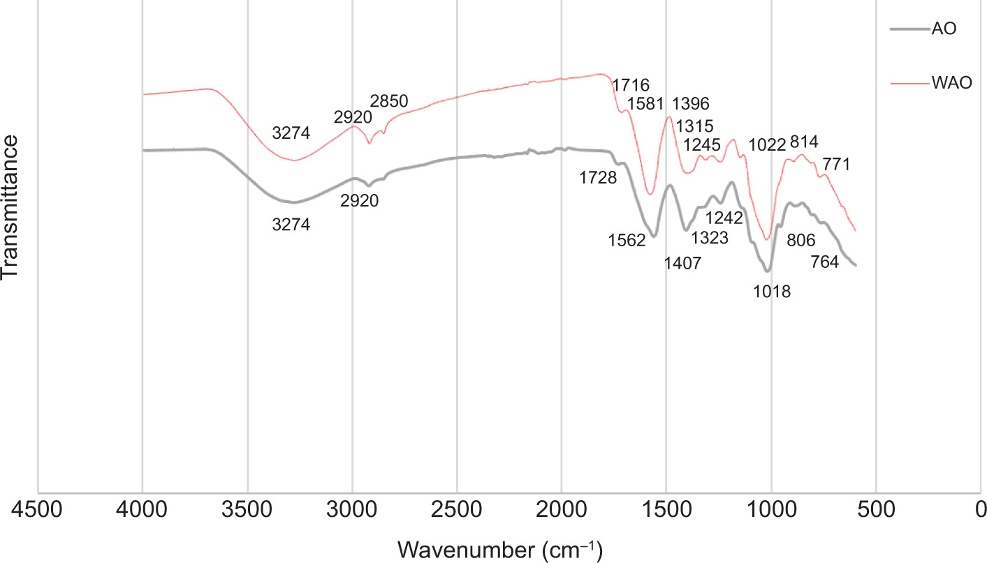
The FTIR spectrometer generates a characteristic spectrum for the compounds known as “fingerprint region” (Kassem et al., 2023). The FTIR spectra of aloe samples are shown in Figure 1 and Table 3. The broad spectrum at 3,274 cm-1 detected for AO and WAO was indicated as a hydroxyl group (-OH), usually because of medicinal compounds, such as alcohols, phenols, and acids and their derivatives. The stretching vibrations of hydroxyl group are shown at the widest wave numbers of 2,900–3,600 cm-1 (Abbasi et al., 2020). The sharp absorption peaks on AO spectra at 2,920 cm-1 and 2,854 cm-1 and on WAO spectra at 2,920 cm-1 and 2,850 cm-1 showed alkyl functional groups. Alkyl functional groups of compounds, such as alkanes and acetates, from aloe buds are observed at 2,855 cm-1 and 2,924 cm-1 (Abbasi et al., 2020). The spectra at 1,728 cm-1 for AO and 1,716 cm-1 for WAO were related to the carbonyl group (C=O). The carbonyl group of acids, aldehydes, and ketones for aloe buds is detected at 1,711 cm-1 (Abbasi et al., 2020). Sharp peaks at 1,562 cm-1 and 1,581 cm-1 on AO and WAO probably contributed to C=C group from aromatic compounds. According to Abbasi et al. (2020), C=C group from aromatic compounds is detected at 1,531 cm-1 and 1601 cm-1 for aloe buds. Absorption at 1,407 cm-1 and 1,396 cm-1 were related to carboxyl group (R-COOH).
Figure 1. The FTIR spectra of Nglipar’s aloe gel (AO) and whole plant (WAO).
Table 3. The FTIR spectral positions and its expected functional groups of Nglipar aloe gel (AO) and whole plant (WAO) (Abbasi et al., 2020; Fardsadegh and Jafarizadeh-Malmiri, 2019).
| FTIR spectral position (cm–1) | Expecting functional group | ||
|---|---|---|---|
| AO | WAO | References | |
| 3,274 | 3,274 | 2,900–3,600 | Hydroxyl group, OH |
| 2,920 | 2,920 | 2,924 | Alkyl compounds, such as alkanes, acetates, esters, acids, ethers, etc. |
| 2,854 | 2,850 | 2,855 | Alkyl compounds, such as alkanes, acetates, esters, acids, and ethers |
| 1,728 | 1,716 | 1,711 | Carbonyl group, C=O, belonged to acids, aldehydes, and ketones |
| 1,562 | 1,581 | 1,531 and 1,601 | C=C belonged to aromatic medicinal compounds |
| 1,407 | 1,396 | 1,402 | Carboxyl group, R-COOH |
| 1,323 | 1,315 | 1,311 | R-COCH3containing compounds and their derivatives |
| 1,242 | 1,245 | 1,232 | R=C-O-C belonged to ethers |
| 1,018 | 1,022 | 1,058 | C=C related to unsaturated five- or six-member ring compounds |
| 806 | 814 | 819 | Alkenes |
| 764 | 771 | 769 | C-H peak belonged to five-member aromatics |
Note: FTIR: Fourier transform infrared spectroscopy.
Carboxyl group from WAO extract was detected at 1,402 cm-1 (Fardsadegh and Jafarizadeh-Malmiri 2019). The spectrum at 1,323 cm-1 for AO and 1,315 cm-1 for WAO matched the functional group, R-COCH3 of the spectrum at 1,311 cm-1 as stated by Abbasi et al. (2020). Spectra at 1,242 cm-1 for AO and 1,245 cm-1 for WAO were similar to those at 1,232 cm-1 related to the ether group, R=C-O-C as shown by Abbasi et al. (2020). The C=C group is related to unsaturated five- or six-member ring compounds, which were probably detected at 1,018 cm-1 for AO and 1,022 cm-1 for WAO. The suggested compound group was detected at 1,058 cm-1 by Abbasi et al. (2020). Spectra at 806 cm-1 and 814 cm-1, detected for AO and WAO, respectively, probably belonged to alkenes. According to Abbasi et al. (2020), alkenes were detected around 819 cm-1. The spectrum at 764 cm-1 on AO and 771 cm-1 on WAO were similar to the peak of 769 cm-1 as shown by Abbasi et al. (2020), which matched the C-H peak belonging to five-member aromatics. Fingerprint region of functional groups is used to assess the quality of a material by comparing it to reference spectra of the same material from different sources (Kassem et al., 2023). Through the fingerprint region of functional groups using FTIR data, we demonstrated that aloe vera in this study was of similar quality as that from other sources.
The LC-HRMS data of aloe vera
The LC-HRMS data detected several active compounds in Nglipar aloe, including polysaccharides, terpenoids, phenolics, and flavonoids (Table 4). Aloesin was detected in both AO and WAO of Nglipar aloe. Nevertheless, aloesin discovered in AO was less than that in WAO. This finding was similar to Añibarro-Ortega et al.’s (2019) results regarding aloesin in AO. The area under curve (AUC) of most terpenoid, phenolic, and flavonoid compounds was lower in AO than in WAO. As reported previously, kaempferol was detected in the peel of Chile aloe but not in its gel (Quispe et al., 2018). Compared to Chile aloe, kaempferol was discovered in Nglipar AO. Glucomannan, galactomannan, and acemannan polysaccharides were also discovered in the Nglipar aloe. Interestingly, acemannan was higher in AO than in WAO. Acemannan is used as a specific marker for aloe polysaccharides (Liu et al., 2019; Metcalfe, 2019).
Table 4. Compounds discovered in aloe gel (AO) and whole plant (WAO) leaves according to LC-HRMS data.
| Compounds | Retention Time (min) | Area under curve (AUC, ×106) | |
|---|---|---|---|
| AO | WAO | ||
| Glucomannan | 1.176 | 6.95 | 9.13 |
| Galactomannan | 1.182 | 13.63 | 33.18 |
| Acemannan | 1.202 | 28.85 | 25.4 |
| Aloesin | 5.871 | 0.67 | 3.82 |
| Kaempferol | 5.916 | 0.01 | 7.16 |
| Gambiriin A1 | 7.985 | 0.02 | 7.52 |
| Eupatorin | 8.033 | 7.57 | 66.63 |
| Curcumene | 8.685 | 0.05 | 1.7 |
| Aloin | 8.856 | – | 372.49 |
| Genistein | 9.271 | 0.09 | 5.8 |
| Dammarenediol | 18.102 | 0.18 | 55.7 |
| Sakuranin | 18.102 | 1.3 | 8.36 |
| Squalene (C30H50) | 20.013 | 5.77 | 1.73 |
Note: LC-HRMS: liquid chromatography high-resolution mass spectrometry.
Aloin is supposed to inducing DNA damage and having carcinogenic effects. Removal of aloin reduces adverse effects and escalate the safety of oral products (Ding et al., 2014; Kim et al., 2023). The result shows high levels of aloin content in WAO but not in AO of Nglipar aloe (Table 4).
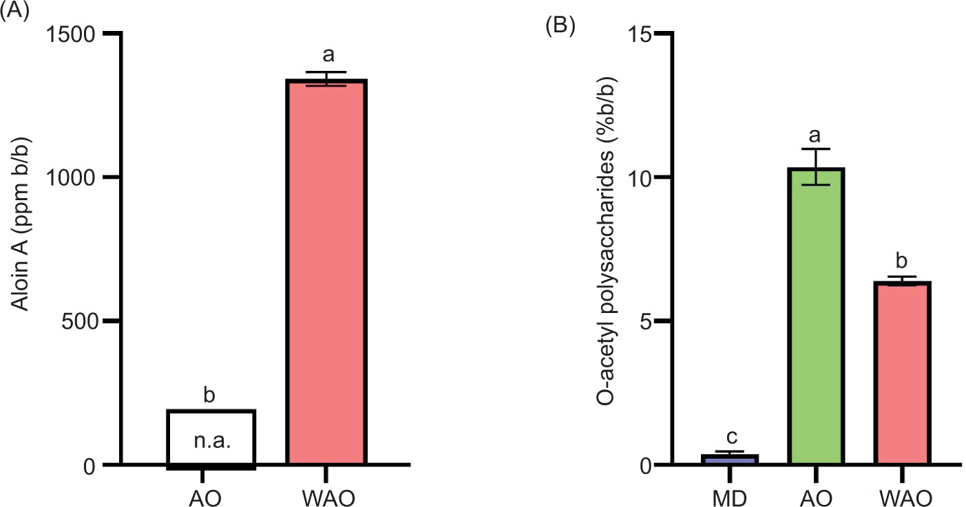
Quantification of aloin A by HPLC
Quantification of aloin A in AO and WAO using HPLC showed that aloin A was not discovered in Nglipar AO but was abundantly found in WAO (Figure 2A). This finding was similar to the LC-HRMS results of aloin (Table 4). Thus, according to the level of aloin in AO, it was considered safe for oral consumption.
Figure 2. Concentration of (A) aloin A and (B) total o-acetyl polysaccharides content in aloe gel (AO) and whole plant (WAO) leaves. The concentration of aloin A was measured using HPLC. The concentration of o-acetyl polysaccharides was measured by UV-Vis spectrophotometer using maltodextrin (MD) as a negative control. Different alphabets show significant differences between samples (P < 0.05).
Measurement of O-acetyl polysaccharides
Because acemannan can be used as a specific marker for aloe polysaccharides (Liu et al., 2019; Metcalfe, 2019), this study calculated the concentration of acemannan by measuring the content of o-acetyl polysaccharides according to Metcalfe (2019). The content of o-acetyl polysaccharides in AO was higher than that in WAO (Figure 2B). This result was consistent with the AUC measurement of acemannan in LC-HRMS results (Table 4). Combining of polysaccharides and aloesin has been reported to possess hepatoprotective activity through antioxidant mechanisms (Yimam et al., 2016). This study revealed that skin removal of aloe leaves drastically reduced the concentration of several active compounds, including aloesin (Table 4). Thus, the in vitro hepatoprotective evaluation was conducted to confirm whether this condition affected the hepatoprotective activity of AO, compared to WAO.
Cell viability
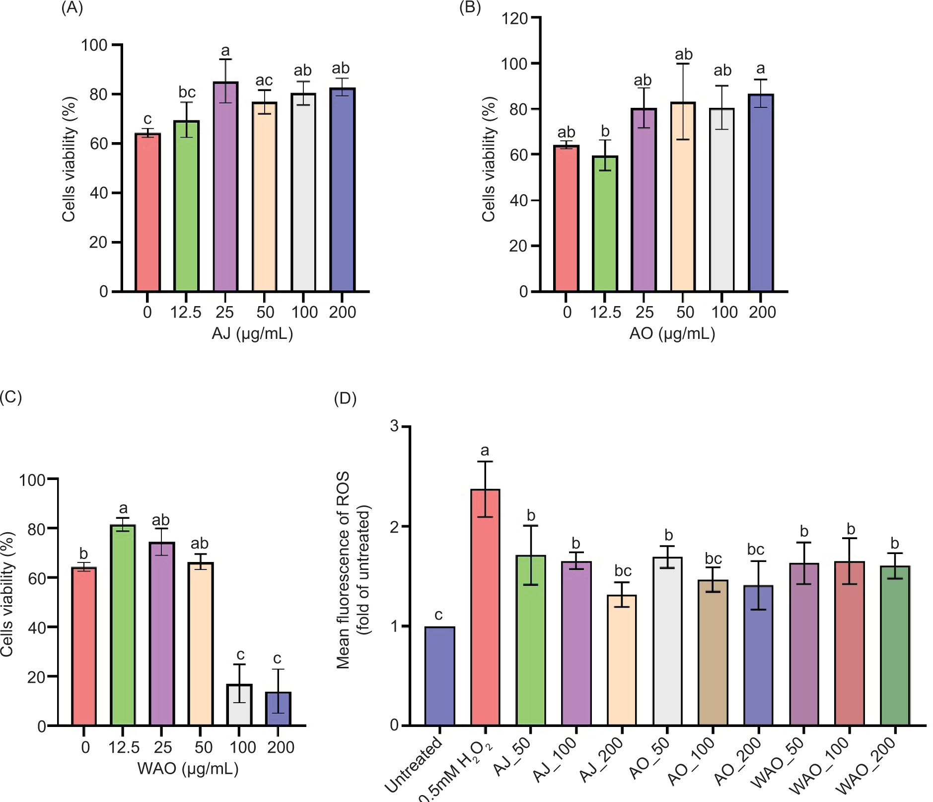
The viability of HepG2 cells was measured by MTT assay. Cell viability was used as an indicator of cytotoxicity prevention. Treatment with H2O2 was used to induce cytotoxicity in HepG2 cells. The result showed that 1.5 mM H2O2 reduced the viability of HepG2 cells by 35.74±1.75%. AJ was used for AO control without drying. Different concentrations of AJ (25, 100, and 200 μg/mL) significantly prevented the cytotoxicity of HepG2 cells by 20.97±8.87%, 16.11±4.80%, and 18.57±3.50%, respectively, after H2O2 treatment (Figure 3A). Different concentrations of AO prevented the cytotoxicity of HepG2 cells after H2O2 treatment, with no significant differences. However, 200 μg/mL of AO showed the highest prevention of 22.44±6.04% (Figure 3B). The lower concentration of WAO (12.5 μg/mL) prevented the cytotoxicity of HepG2 cells by 17.20±2.70% after H2O2 treatment. Nevertheless, the higher concentration of WAO tended to induce cytotoxicity of HepG2 cells more than that by H2O2 treatment (Figure 3C). The result revealed that treatment with AO extract prevented H2O2-induced cytotoxicity in HepG2 cells, compared to WAO.
Figure 3. Hepatoprotective activity of Nglipar AJ, AO, and WAO. Cell viability by varying concentrations (μg/mL) of (A) AJ, (B) AO, and (C) WAO on HepG2 cells after H2O2 induction. (D) Intracellular ROS levels of HepG2 cells after extract treatment (μg/mL). All measurements are analyzed according to the methods. Different alphabets show significant differences among samples (P < 0.05).
Measurement of reactive oxygen species level
Alterations in ROS at intracellular level was monitored using the DCFDA staining ROS-based assay. The ROS of cells was measured to show the level of oxidative stress after sample treatment. The result showed that H2O2 treatment increased ROS level by 2.4-fold, compared to untreated cells, while the ROS level of all aloe extract treatments was lower than that of H2O2 treatment. AJ, 200 μg/mL, and 100 and 200 μg/mL of AO showed no significant ROS level, compared to untreated cells (Figure 3D).
Docking of metabolite compound of aloe gel against TACE protein
As a main protein having a role in fibrogenesis and other liver injury models, high expression of TNF-α in inflammation cases was caused by the increased level of TACE. Thus, inhibition of TACE may decrease hepatic fibrosis through TNF-α signal pathway (Lakshmi et al., 2019). Studies about the affinity of different compounds discovered in AO (Table 4) toward TACE enzyme were limited, especially for the in silico docking method. In this study, various compounds discovered in LC-HRMS data (Table 4) were tested for in silico docking test of TACE enzyme. Polysaccharides were excluded from this test because the software cannot process the high molecular weights of these compounds.
The docking protocol in this study was valid because the RMSD of TACE (2VF5) conforms to the required value (≤2 Å) after running with the docking protocol. Internal native ligand (IK682) and silibinin were used as reference standards (Mogadem et al., 2022), and 13 compounds detected in aloe gel were tested for its binding affinity against TACE enzyme using this protocol. The docking score was measured as a parameter for protein–ligand interaction. The native ligand showed the lowest docking score (-124.5±5.05). The other 14 compounds (including positive TACE inhibitor silibinin) that showed interaction with TACE enzyme from the lowest to the highest docking scores were squalene (C30H50), gambiriin A1, silibinin, naringenin 4’ methoxy 7 o-glucuronide, sakuranin, sakuranetin, curcumene, aloesin, 7 methyl ether 2’ feruloylaloesin, isoaloeresin D, dammarenediol, genistein, kaempferol, and eupatorin (Table 5). Squalene and gambiriin A1 had a lower docking score than the reference standard silibinin, while other compounds showed higher docking scores.
Table 5. Protein–ligand interaction of TACE protein (2VF5) with expected compounds in aloe leaves.
| No. | Compound | Docking score | H-bond | Hydrophobic bond | External bond | |
|---|---|---|---|---|---|---|
| Residue | Distance (Å) | |||||
| 1. | IK682 (native ligand) | –124.5±5.05 | Zn | 2.20; 2.37 | Ala439; Asn447; Gly346; Leu350; Leu401; Pro437; Ser441; Thr347; Tyr433; Tyr436; Val402; Val434; Val440 | – |
| 3.34 | ||||||
| 3.13 | ||||||
| 3.18 | ||||||
| 2.86 | ||||||
| 2. | Silibinin(TACE inhibitor) | –99.2±1.25 | Ala439; Ile438; Glu398; Glu406; Gly346; Leu348; Leu401; Met435; Pro437; Val402 | His405 (2); Val434 (1) | ||
| 3. | 7 Methyl ether 2’-feruloylaloesin | –88.7±1.15 | His405; His415; Ile438; Glu406; Gly349; Leu401; Met435; Pro437; Ser441; Tyr433; Tyr436; Val402 | Ala439 (5); Glu398 (1); Val434 (4) | ||
| 4. | Aloesin | –89.2±2.07 | Zn | 2.79 | Ile438; Glu406; Gly349; Leu348; Met435; Pro437; Thr347; Tyr433; Val402; Val 434 | Ala439 (1); His405 (1); Tyr436 (1) |
| 5. | Isoaloeresin D | –88.8±2.11 | His405 | 3.09 | His415; Ile438; Glu398; Glu406; Gly349; Leu348; Leu350; Leu401; Met435; Ser441; Tyr436; Val402; Val440 | Ala439 (3); Pro437 (1); Val434 (3) |
| 6. | Naringenin 4’-Methoxy 7 o-glucuronide | –93.3±3.39 | His405 | 2.89 | Ala439; Ile438; Leu348; Leu401; Met435; Pro437; Tyr433; Val402 | Glu406 (3); Gly349 (1); Val434 (1) |
| 2.37 | ||||||
| 7. | Eupatorin | -80.4±1.83 | Gly349 | 2.36 | – | – |
| Leu348 | 2.18 | |||||
| 8. | Curcumene | –89.8±0.39 | His405; Glu406; Gly349; Leu348; Thr347; Tyr436; Val402; Val440 | Ala439 (1); Ile438 (1) | ||
| 9. | Dammarenediol | –88.9±0.59 | His405 | 3.21 | Ala439; Asn447; Ile438; Glu406; Gly346; Gly349; Gly442; Leu348; Leu401; Pro437; Ser441; Thr347; Tyr433; Val402; Val434 | |
| 10. | Gambiriin A1 | –102.8±2.86 | His409 | 2.41 | His415; Ile438; Val440 | Ala439 (7); His405 (1); Ile438 (2); Leu348 (1); Leu401 (1); Pro437 (1); Val434 (2) |
| Glu406 | 3.16 | |||||
| 11. | Genistein | –86.8±0.64 | Thr347 | 3.04 | Ala439; Gly349; Leu348; Pro437; Tyr436; Val434 | Thr347 (1) |
| 12. | Kaempferol | –82.2±0.55 | Ala439; Ile438; Glu406; Gly349; Leu348; Pro437; Thr347; Tyr436; Val402; Val440 | |||
| 13. | Sakuranetin | –90.7±2.14 | Ala439; His405; Ile438; Glu406; Gly349; Leu348; Pro437; Thr347; Tyr436; Val402; Val440 | |||
| 14. | Sakuranin | –92.5±0.79 | Zn | 2.57 | Ile438; Glu398; Leu348; Lys448; Pro437; Val402; Val434 | Ala439 (3); Asn447 (1); Leu401 (4) |
| His405 | 2.33 | |||||
| Thr347 | 2.06 | |||||
| 15. | Squalene | –116.6±2.54 | Gly346; Leu348; Leu350; Leu401; Met435; Pro437; Thr347; Thr403; Thr404; Tyr433; Tyr436; Val402; Val440 | Ala439 (1); His405 (5); Ile438 (3); Gly349 (1); Val434 (2) | ||
Note: TACE: tumor necrosis factor (TNF)-α converting enzyme.
Native inhibitor of TACE protein has active sites on Glu406, Gly349, and Leu348, while silimaryn/silybinin has active sites on Glu406, Gly349, Glu398, Asn447, Tyr433, and Lys432 (Borah et al., 2016). The TACE–ligand interaction in this study showed that the native ligand of TACE (IK682) had six hydrogen bonds and 13 hydrophobic interactions, including its interaction with active sites Gly349, Leu348, Asn447, and Tyr433. Silibinin had 10 hydrophobic interactions and three external bonds, including its interaction with active sites Glu406, Glu398, and Leu348. Squalene had 13 hydrophobic interactions and 12 external bonds, including its interaction with active sites Gly349, Leu348, and Tyr433. Meanwhile, gambiriin A1 had two hydrogen bonds, three hydrophobic interactions, and 15 external bonds, including its interaction with active site Glu406 (Table 5). Nevertheless, some other compounds also interacted with the active sites of TACE protein while the docking score was still higher than that of the reference ligand.
Aloesin and its derivatives have various beneficial bioactivities for health (Handayani et al., 2021, 2023). In this study, aloesin was rich in WAO but less in AO (Table 4). On the other hand, AO contained higher o-acetyl polysaccharides than WAO (Figure 2B). Polysaccharides have a role in hepatoprotective effect (Qu et al., 2020). Combining polysaccharides and aloesin increases hepatoprotective activity through an antioxidant mechanism (Yimam et al., 2016). Interestingly, the data of viability cells revealed that AO inhibited the cytotoxicity of HepG2 cells after H2O2 treatment. Meanwhile, the higher concentration of WAO tended to induce cytotoxicity in HepG2 cells (Figures 3A–C). The cytotoxicity of WAO at high concentrations probably was due to the high presence of aloin (Figure 2A). WAO contained more aloin and other anthraquinones than AO. Administration of commercial AO beverages containing less aloin (3.43 ppm) for 90 days did not alter the histopathology of mice’s organs (Hayes et al., 2024). Aloin at a low concentration protects SH-SY5Y cells against peroxide-induced toxicity, but not at a higher concentration (Kaparakou et al., 2021).
One of the hepatoprotective mechanisms of aloe is the prevention of ROS production (Handayani et al., 2021). The measurement of ROS revealed that treatment with AO or WAO after H2O2 treatment inhibited ROS levels, compared to H2O2 treatment alone. AJ was used to ensure that the AO drying method did not affect AO extract’s bioactivity. The result showed that the ROS level of AJ and AO did not differ significantly. Thus, the drying method in this study did not affect the bioactivity of AO extract. The insignificant ROS levels of 200 μg/mL for AJ, and 100 μg/mL and 200 μg/mL for AO, compared to untreated cells, revealed their prevention activity toward H2O2-induced ROS production (Figure 3D). Thus, the hepatoprotective mechanism of AO revealed in this study was due to the prevention of ROS production. Pro-inflammatory cytokine, TNF-α, induces ROS production (Wu and Pan 2019).
Aloe vera inhibited TNF-α and did not alter the histopathological pattern of mice liver (Gupta et al., 2023). High expression of TNF-α in inflammation is caused by the increased TACE level. TACE, also known as metalloproteinase 17 (ADAM17), has an important role in the cleavage and release of TNF-α. Soluble TNF-α binds to its receptors and initiates downstream signaling pathways. Increasing TACE activity in oxidative stress induces insulin resistance and hepatitis. Thus, the strategies targeting TACE are considered effective in treating liver diseases via TNF-α signaling pathway (Lakshmi et al., 2019; Srinivas et al., 2024). The affinity of all compounds toward TACE receptor was less than that of IK682 (native ligand). Interestingly, the affinity of squalene and gambiriin A1 discovered in Nglipar aloe was higher than the reference compound silibinin (Table 5). Silymarin and silibinin are the known compounds that act as TACE inhibitors (Borah et al., 2016). Squalene is generally discovered in sea fish. Nevertheless, squalene is also discovered in various plants. A high concentration of squalene can be extracted from tea leaves also (Sheng et al., 2022). According to LC-HRMS data, squalene showed a bigger AUC in AO than WAO (Table 4). On the other hand, even though gambiriin A1 was first found in Uncaria gambir Roxb, this compound was also found in other plants (Mazlan et al., 2018; Zhou et al., 2022). Thus, the affinity of squalene and gambiriin A1 toward TACE enzyme probably acted as TACE inhibitor.
In addition, Van der Waals (VdW) forces are an essential parameter for the molecular recognition of a ligand by the target receptor-binding pocket (Bitencourt-Ferreira et al., 2019). The ligplot software showed all external bonds, including VdW forces. In this study, silibinin, squalene, and gambiriin A1, and not other compounds, had external bonds with His405 and Val434 (Table 5). His405, His409, and His415 had catalytic interaction with Zn, followed by activation of TACE enzyme (Hermenean et al., 2017). Squalene had five external bonds with His405, while gambiriin A showed hydrogen interaction with His409, a hydrophobic bond with His415, and one external bond with His405. These interactions could increase the affinity of squalene and gambiriin A1 toward TACE protein and break the catalytic interaction of amino acid residues of TACE with Zn. Thus, squalene and gambiriin A1 probably had a role in the hepatoprotective effects of AO through inhibition of TACE enzyme. The data support the hepatoprotective activity and the safety of AO, especially from Nglipar plantation in Indonesia.
Conclusion
This study of aloe vera plantation Nglipar in Indonesia demonstrated that AO provided a higher protection than WAO against H2O2-induced hepatotoxicity in HepG2 cells. Soil analyses from four sites around Nglipar revealed clay, silty clay, and loamy textures, with no detectable levels of heavy metals (Hg, As, or Pb), suggesting a favorable environment condition for growth of aloe vera. The functional groups of both AO and WAO were similar; however, aloin A was abundant in WAO but absent in AO extract. AO extract effectively inihibited H2O2-induced toxicity in HepG2 cells, whereas WAO offered protection only at lower concentrations and caused toxicity at higher concentrations. Both 100 μg/mL and 200 μg/mL of AO extract significantly reduced H2O2-induced ROS production. LC-HRMS analysis revealed that squalene was more concentrated in AO than in WAO, and docking studies revealed that squalene had the highest affinity for TACE protein. These findings confirm that AO extract has potential inhibitory activity against oxidative stress-induced damage in HepG2 cells, suggesting it can serve as a safe and strong hepatoprotective agent. However, this study has several limitations that must be addressed in the future research. First, the hepatoprotective effects of aloe extracts were investigated only in HepG2 cells. Additionally, H2O2 was the sole oxidative stress inducer used, which limits the assessment of protective effects. Therefore, the future studies must include animal models and multiple hepatotoxic agents to confirm comprehensively the hepatoprotective activity.
Data Availability
The data supporting the findings are available upon reasonable request.
Author Contributions
Sari Haryanti: formal analysis, investigation, data curation, writing – original draft, and writing – review and editing. Mujiyanto Mujiyanto: investigation, data curation, writing – original draft, and visualization. Nur Maulidah Rahmah: data curation, visualization, and writing – review & editing. Ade Erma Suryani, Santosh Chokkakulula, Khoirun Nisa, Balasubramani Ravindran, and Soon Woong Chang: formal analysis, and writing – review and editing. Sri Handayani and Ravishankar Ram Mani: conceptualization, methodology, investigation, resources, data curation, writing – review and editing, visualization, supervision, and project administration.
Conflicts of Interest
The authors declared no conflict of interest.
Funding
This work was supported by Prioritas Nasional of Deputi Ilmu Pengetahuan Hayati, Indonesian Institute of Sciences (LIPI) (Grant No. B-10405/IPH/HK.01.03/XI/2020) and PROGRAM RISET DAN INOVASI UNTUK INDONESIA MAJU (RIIM Kompetisi) funding from the Indonesia Endowment Fund for Education Agency, Ministry of Finance of the Republic of Indonesia, and National Research and Innovation Agency of Indonesia (BRIN) (Grant No. 60/II/HK/2022). We are thankful for the support provided by UCSI University for the support provided by the Center of Excellence for Research, Value Innovation and Entrepreneurship (CERVIE) and Research Excellence and Innovation Grant (REIG) with code REIG-FPS-2025/038.
REFERENCES
Abbasi, Muhammad S.A., Tahir, M.S., and Meer, S. 2020. FTIR spectroscopic study of aloe vera Barbadensis Mill buds. Asian Journal of Chemical Sciences 7(4): 1–6. 10.9734/ajocs/2020/v7i419026
Abdelhak, M. 2022. Soil improvement in arid and semiarid regions for sustainable development. In: Jhariya, M.K., Meena, R.S., Banerjee, A., and Meena, S.N. (eds.) Natural Resources Conservation and Advances for Sustainability. Elsevier, Amsterdam, The Netherlands, Chap. 4, pp. 73–90.
Adlakha, K., Koul, B., and Kumar, A. 2022. Value-added products of aloe species: panacea to several maladies. South African Journal of Botany 147: 1124–1135. 10.1016/j.sajb.2020.12.025
Akbari, B., Baghaei-Yazdi, N., Bahmaie, M., and Abhari, F.M. 2022. The role of plant-derived natural antioxidants in reduction of oxidative stress. BioFactors 48(3): 611–633. 10.1002/biof.1831
Al-Sheddi, Ebtesam S., Farshori, Nida N., Al-Oqail, Mai M., Alblwi, F., Ahmad, J., Al-Khedhairy, Abdulaziz A., and Siddiqui, Maqsood A. 2024. Hepatoprotective effect of date fruit extract against ethanol-induced apoptosis in human hepatoma (HepG2) cells. Tissue and Cell 90: 102519. 10.1016/j.tice.2024.102519
Añibarro-Ortega, M., Pinela, J., Barros, L., Ćirić, A., Silva, Soraia P., Coelho, E., Mocan, A., Calhelha, Ricardo C., Soković, M., Coimbra, M.A., and Ferreira, Isabel C.F.R. 2019. Compositional features and bioactive properties of aloe vera leaf (fillet, mucilage, and rind) and flower. Antioxidants 8(10): 444. 10.3390/antiox8100444
Artanti, N., Dewijanti, I.D., Muzdalifah, D., Windarsih, A., Suratno, S., and Handayani, S. 2023. Alpha-glucosidase inhibitory activity of the combination of Caesalpinia Sappan L., and Garcinia mangostana extract. Journal of Applied Pharmaceutical Science 13(5): 189–198. 10.7324/JAPS.2023.117478
Babu, S.N., and Noor, A. 2021. Bioactive constituents of the genus aloe and their potential therapeutic and pharmacological applications: a review. Journal of Applied Pharmaceutical Sciences 10(11): 133–145. 10.7324/JAPS.2020.101118
Bitencourt-Ferreira, G., Veit-Acosta, M., and Filgueira de Azevedo, W. 2019. Van der Waals potential in protein complexes. Methods in Molecular Biology 2053: 79–91. 10.1007/978-1-4939-9752-7_6
Borah, Pallab Kumar, Chakraborty, S., Jha, Anupam N., Rajkhowa, S., and Duary, R.K. 2016. In silico approaches and proportional odds model towards identifying selective ADAM17 inhibitors from anti-inflammatory natural molecules. Journal of Molecular Graphics & Modelling 70: 129–139. 10.1016/j.jmgm.2016.10.003
BPS Kab.Gunungkidul. 2019. Kecamatan Nglipar Dalam Angka 2019. Badan Pusat Statistik Kabupaten Gunungkidul, Indonesia. Available at: https://gunungkidulkab.bps.go.id/publication/2019/09/26/9f287b34d06165a6f6658d90/kecamatan-nglipar-dalam-angka-2019.html (Accessed 16/8/2024).
BPS Yogyakarta. 2020. Jumlah Curah Hujan dan Hari Hujan Menurut Bulan di D.I. Yogyakarta, 2019 – Tabel Statistik. Central Statistics Agency (BPS), Indonesia. Available at: https://yogyakarta.bps.go.id/id/statistics-table/1/OTMjMQ==/jumlah- (Accessed 12/9/2024).
Chaudhary, P., Janmeda, P., Docea, A.O., Yeskaliyeva, B., Abdull Razis, A.F., Modu, B., Calina, D., and Sharifi-Rad, J. 2023. Oxidative stress, free radicals and antioxidants: potential crosstalk in the pathophysiology of human diseases. Frontiers in Chemistry 11: 1158198. 10.3389/fchem.2023.1158198
Chinnadurai, A., Jesuraj, S., Eswaran, K., Doraiswamy, U., Nathan, B., and Sundar, B. 2024. Aloesin unveiled: molecular docking investigation of aloe vera as anti-inflammatory activity against tumour necrosis factor alpha. Indian Journal of Pharmaceutical Sciences 86(5): 1756–1764. 10.36468/pharmaceutical-sciences.1442
Chowdhury, T., Rahman, Md A., Nahar, K., Chowdhury, Md A.H., and Khan, Md S.I. 2018. Growth and yield performance of aloe vera grown in different soil types of Bangladesh: yield performance of aloe vera in different soils. Journal of the Bangladesh Agricultural University 16(3): 448–456. 10.3329/jbau.v16i3.39416
Ding, W.-J., Wu, X.-F., Zhong, J.-S., and Wan, J.-Z. 2014. Effects of temperature, pH and light on the stability of aloin A and characterisation of its major degradation products. International Journal of Food Science & Technology 49(7): 1773–1779. 10.1111/ijfs.12500
Dobbins, Dylan C., Marcelo-Silva, J., and Siebert, S.J. 2021. Screening the phytoextractability of trace metals by aloe cryptopoda baker and aloe vera (L.) burm. f. cultivated on mine tailings. South African Journal of Botany 140: 110–113. 10.1016/j.sajb.2021.03.042
EFSA Panel on Food Additives and Nutrient Sources added to Food (ANS), Younes, M., Aggett, P., Aguilar, F., Crebelli, R., Filipič, M., Frutos, M.J., et al. 2018. Safety of hydroxyanthracene derivatives for use in food. EFSA Journal 16(1): e05090. 10.2903/j.efsa.2018.5090
Fardsadegh, B., and Jafarizadeh-Malmiri, H. 2019. Aloe vera leaf extract mediated green synthesis of selenium nanoparticles and assessment of their in vitro antimicrobial activity against spoilage fungi and pathogenic bacteria strains. Green Processing and Synthesis 8(1): 399–407. 10.1515/gps-2019-0007
García, D., Perdomo, M.E., and Magomedov, I. 2023. Study case: correlational analysis of pH and weekly aloe growth. BIO Web of Conferences 63: 7007. 10.1051/bioconf/20236307007
Guo, X., and Mei, N. 2016. Aloe vera: a review of toxicity and adverse clinical effects. Journal of Environmental Science and Health. Part C: Environmental Carcinogenesis & Ecotoxicology Reviews 34(2): 77–96. 10.1080/10590501.2016.1166826
Gupta, V.K., Park, U., Siddiqi, N.J., Huh, Y.S., and Sharma, B. 2023. Amelioration of hepatotoxic and neurotoxic effect of cartap by aloe vera in Wistar rats. Toxics 11(5): 472. 10.3390/toxics11050472
Gupta, V.K., Siddiqi, Nikhat J., Ojha, A.K., and Sharma, B. 2019. Hepatoprotective effect of aloe vera against cartap-and malathion-induced toxicity in Wistar rats. Journal of Cellular Physiology 234(10): 18329–18343. 10.1002/jcp.28466
Handayani, S., Aprilia, D., Nisa, K., Rosyida, V.T., Wulanjati, M.P., Windarsih, A., Darsih, C., Frediansyah, A., and Haryanti, S. 2021. A mini-review: possible mechanisms of hepatoprotective effect of aloe vera gel. Indonesian Journal of Cancer Chemoprevention 12(3): 170–179. 10.14499/indonesianjcanchemoprev12iss3pp170-179
Handayani, S., Ni’maturrohmah, D., Indrianingsih, A.W., Nisa, K., Windarsih, A., Darsih, C., Sefrienda, A.R., Suryani, A.E., and Haryanti, S. 2023. Molecular docking study of aloesin and its derivatives as potential antiaging agents. In: Proceedings of the 1st International Conference for Health Research – BRIN (ICHR 2022); Advances in Health Sciences Research. Atlantis Press, Dordrecht, The Netherlands, pp. 288–299. 10.2991/978-94-6463-112-8_28
Hayes, A., Wallace, P.P., Clemens, R., Singer, A.W., and Bauter, M.R. 2024. Evaluation of 90-day repeated dose oral toxicity of an aloe vera inner leaf gel beverage. Food and Chemical Toxicology 189: 114726. 10.1016/j.fct.2024.114726
Hermenean, A., Mariasiu, T., Navarro-González, I., Vegara-Meseguer, J., Miuțescu, E., Chakraborty, S., and Pérez-Sánchez, H. 2017. Hepatoprotective activity of chrysin is mediated through TNF-α in chemically induced acute liver damage: an in vivo study and molecular modeling. Experimental and Therapeutic Medicine 13(5): 1671–1680. 10.3892/etm.2017.4181
Hu, Y., Xiang, X., Zhang, Y., Tian, Z., and Wang, L. 2022. Aloin promotes oral squamous cell carcinoma cell apoptosis and autophagy through Akt/mTOR pathway. Quality Assurance and Safety of Crops & Foods 14(2): 58–65. 10.15586/qas.v14i2.978
Kaloni, D., Tiwari, A., and Biswas, S. 2019. Aloe vera as an antagonist for TNF-Α: In-silico study. International Journal of Innovative Science and Research Technology 4(10): 78–83.
Kaparakou, E.H., Kanakis, C.D., Gerogianni, M., Maniati, M., Vekrellis, K., Skotti, E., and Tarantilis, P.A. 2021. Quantitative determination of aloin, antioxidant activity, and toxicity of leaf gel products from Greece. Journal of the Science of Food and Agriculture 101(2): 414–423. 10.1002/jsfa.10650
Kassem, A., Abbas, L., Coutinho, O., Opara, S., Najaf, H., Kasperek, D., Pokhrel, K., Li, X., and Tiquia-Arashiro, S. 2023. Applications of Fourier transform-infrared spectroscopy in microbial cell biology and environmental microbiology: advances, challenges, and future perspectives. Frontiers in Microbiology 14: 1304081. 10.3389/fmicb.2023.1304081
Kim, S-T., Pressman, P., Clemens, R., Moore, A., Hamilton, R., and Wallace Hayes, A. 2023. The absence of genotoxicity of aloe vera beverages: a review of the literature. Food and Chemical Toxicology 174: 113628. 10.1016/j.fct.2023.113628
Lakshmi, C., Nair, S., Balachandran, S., Arul, D.D., Ronaldo, A.A., and Hubert, J.I. 2019. DFT Analysis on spectral and NLO Properties of (2E)-3-[4-(dimethylamino) phenyl]-1-(naphthalen-2-Yl) prop-2-en-1-one; a d-π-A chalcone derivative and its docking studies as a potent hepatoprotective agent. Chemical Data Collections 20: 100205. 10.1016/j.cdc.2019.100205
Lee, J.Y., Kim, H., Jeong, Y., and Kang, C.H. 2021. Lactic acid bacteria exert a hepatoprotective effect against ethanol-induced liver injury in HepG2 cells. Microorganisms 9(9): 1844. 10.3390/microorganisms9091844
Lestari, E.N.E., Nisa, K., Suryani, A.E., and Kusumaningsih, T. 2024. Encapsulation of Peperomia pellucida (L.) kunth leaf extract for postharvest preservation of Malang Apple (Malus sylvestris) at ambient storage. Food Bioscience 61: 104808. 10.1016/j.fbio.2024.104808
Liu, C., Cui, Y., Pi, F., Cheng, Y., Guo, Y., and Qian, H. 2019. Extraction, purification, structural characteristics, biological activities and pharmacological applications of acemannan, a polysaccharide from aloe vera: a review. Molecules (Basel, Switzerland) 24(8): 1554. 10.3390/molecules24081554
Mazlan, O., Aizat, W. Mohd., Nataqain Baharum, S., Azizan, K.A., and Noor, N. Mohd. 2018. Metabolomics analysis of developing Garcinia mangostana seed reveals modulated levels of sugars, organic acids and phenylpropanoid compounds. Scientia Horticulturae 233: 323–330. 10.1016/j.scienta.2018.01.061
Meiyanto, E., Putri, D.D.P., Susidarti, R.A., Murwanti, R., Sardjiman, S., Fitriasari, A., Husnaa, U., Purnomo, H., and Kawaichi, M. 2014. Curcumin and its analogues (PGV-0 and PGV-1) enhance sensitivity of resistant MCF-7 cells to doxorubicin through inhibition of HER2 and NF-kB Activation. Asian Pacific Journal of Cancer Prevention 15(1): 179–184. 10.7314/APJCP.2014.15.1.179
Metcalfe, C. 2019. Quantitation of aloe vera polysaccharides by O-acetyl and UV-Vis spectrophotometry: first action 2018.14. Journal of AOAC International 102(4): 1091–1094. 10.5740/jaoacint.18-0400
Mogadem, A., Naqvi, A., Almamary, Mohamed A., Ahmad, W.A., Jemon, K., and El-Alfy, S.H. 2022. Hepatoprotective effects of flexirubin, a novel pigment from Chryseobacterium artocarpi against carbon tetrachloride-induced liver injury: an in vivo study and molecular modeling. Toxicology and Applied Pharmacology 444: 116022. 10.1016/j.taap.2022.116022
Nair, Hema C., Joseph, A., and Gopinathan, V.P. 2018. GIS based landform classification using digital elevation model: a case study from two river basins of southern western ghats, Kerala, India. Modeling Earth Systems and Environment 4(4): 1355–1363. 10.1007/s40808-018-0490-5
Padmanabhan, P., and Jangle, S.N. 2014. Hepatoprotective activity of herbal preparation (Hp-4) against alcohol-induced hepatotoxicity in mice. International Journal of Applied Sciences and Biotechnology 2(1): 50–58. 10.3126/ijasbt.v2i1.9346
Qu, J., Huang, P., Zhang, L., Qiu, Y., Qi, H., Leng, A., and Shang, D. 2020. Hepatoprotective effect of plant polysaccharides from natural resources: a review of the mechanisms and structure-activity relationship. International Journal of Biological Macromolecules 161: 24–34. 10.1016/j.ijbiomac.2020.05.196
Quispe, C., Villalobos, M., Bórquez, J., and Simirgiotis, M. 2018. Chemical composition and antioxidant activity of aloe vera from the pica oasis (Tarapacá, Chile) by UHPLC-Q/Orbitrap/MS/MS. Journal of Chemistry 2018: e6123850. 10.1155/2018/6123850
Sahoo, S., Rath, D., Kar, D.M., and Pattanaik, S. 2023. Hepatoprotective potency of Litsea glutinosa (L.) C.B. rob. leaf methanol extract on H2O2-induced toxicity in HepG2 cells. Journal of Ethnopharmacology 304: 116076. 10.1016/j.jep.2022.116076
Sheng, Y.Y., Xiang, J., Wang, K.R., Li, Z.Y., Li, K., Lu, J.L., Ye, J.H., Liang, Y.R., and Zheng, X.Q. 2022. Extraction of squalene from tea leaves (Camellia sinensis) and its variations with leaf maturity and tea cultivar. Frontiers in Nutrition 9: 755514. https://www.frontiersin.org/articles/10.3389/fnut.2022.755514
Shokri, F., Ziarati, P., and Mousavi, Z. 2016. Removal of selected heavy metals from pharmaceutical effluent by aloe vera L. Biomedical and Pharmacology Journal 9(2): 705–713. 10.13005/bpj/993
Srinivas, A.N., Suresh, D., Vishwanath, P.M., Satish, S., Santhekadur, P.K., Koka, S., and Kumar, D.P. 2024. TACE inhibition: a promising therapeutic intervention against AATF-mediated steatohepatitis to hepatocarcinogenesis. Molecular Oncology 18(8): 1940–1957. 10.1002/1878-0261.13646
Sultana, T., Chowdhury, A.H., Rahman, A., Saha, B.K., Chowdhury, T., Islam, Mohammed A., and Fancy, R. 2021. Phosphorous use efficiency and its requirement for aloe vera cultivated on silty loam soils. Communications in Soil Science and Plant Analysis 52(3): 268–285. 10.1080/00103624.2020.1862152
Suryani, A.E., Nisa, K., Handayani, S., Darsih, C., Wuryastuty, H and Yanuartono, Y. 2025. The UHPLC-HRMS profiling, in vitro-antioxidant and pancreatic lipase inhibitory activities of peronema canescens leaves extract and fractions from Indonesia. Journal of Applied Pharmaceutical Science 15(5): 75–089. 10.7324/JAPS.2025.214965
Wu, L., and Pan, Y. 2019. Reactive oxygen species mediate TNF-α-induced inflammatory response in bone marrow mesenchymal cells. Iranian Journal of Basic Medical Sciences 22(11): 1296–1301. 10.22038/ijbms.2019.37893.9006
Yang, Yu., Wu, J.-j., Xia, J., Wan, Y., Xu, J.-F., Zhang, L., Liu, D., Chen, L., Tang, F., Ao, H., and Peng, C. 2022. Can aloin develop to medicines or healthcare products? Biomedicine & Pharmacotherapy 153: 113421. 10.1016/j.biopha.2022.113421
Yimam, M., Jiao, P., Moore, B., Hong, M., Cleveland, S., Chu, M., Jia, Q., Lee, Y-C., Kim, H-J., Nam, J-B., Kim, M-R., Hyun, E-J., Jung, G., and Do, S.G. 2016. Hepatoprotective activity of herbal composition SAL, a standardize blend comprised of Schisandra chinensis, Artemisia capillaris, and Aloe barbadensis. Journal of Nutrition and Metabolism 2016: 3530971. 10.1155/2016/3530971
Zhou, B., Alania, Y., Reis, M., Jing, S-Xi., McAlpine, James B., Bedran-Russo, Ana K., Chen, S-N., Ferreira, D., and Pauli, G.F. 2022. Seco B-type oligomers from Pinus massoniana expand the procyanidin chemical space and exhibit dental bioactivity. Journal of Natural Products 85(12): 2753–2768. 10.1021/acs.jnatprod.2c00664
Zhou, X., and Zhao, X. 2021. Gastrodin represses hydrogen peroxide-induced oxidative stress in retinal pigment epithelial cells through p38MAPK/iNOS pathway. Quality Assurance and Safety of Crops & Foods 13(4): 24–30. 10.15586/qas.v13i4.969